Alternate header for print version
Advanced search
Contributors
Help
Submit
Search
menu
Cell Process
Cell Component
Cell Type
Organism
Microbial
Alzheimer's
Data Sets
University of California, San Diego
9500 Gilman Drive
La Jolla, CA 92093-0608, USA
Voice
: (858) 534-0276
Fax
: (858) 534-7497
Email
: dorloff@ncmir.ucsd.edu
Grouped images - the images shown below are related
CIL:10970
NCBI Organism Classification
Rhipicephalus appendiculatus
Biological Process
nucleus organization
Cellular Component
nucleus
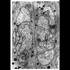
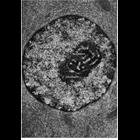
Transmission electron micrograph of the salivary gland of the tick Rhipicelphals...
CIL:10971
NCBI Organism Classification
Homo sapiens
Biological Process
adaptive immune response
Cellular Component
nucleus
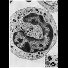
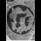
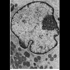
Transmission electron micrograph of a section of a human polymorphonuclear neutr...
CIL:10973
NCBI Organism Classification
Chinchilla
Biological Process
nucleus organization
Cellular Component
nucleus
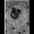
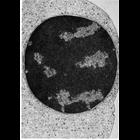
Transmission electron micrograph of a section of Chinchilla epididimus showing t...
CIL:10974
NCBI Organism Classification
none specified
Biological Process
nucleus organization
Cellular Component
nucleus
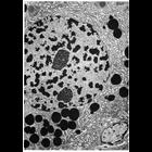
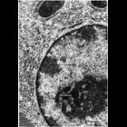
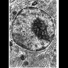
Transmission electron micrograph illustrating the 'classic' appearance of the nu...
CIL:10976
NCBI Organism Classification
Myotis lucifugus
Biological Process
nucleus organization
Cellular Component
nucleus
Transmission electron micrograph of nucleus showing the characteristic features ...
CIL:10977
NCBI Organism Classification
Cavia porcellus
Biological Process
nucleus organization
Cellular Component
nuclear heterochromatin
Transmission electron micrograph showing the extensive accumulation of darkly st...
CIL:10978
NCBI Organism Classification
Cavia porcellus
Biological Process
adaptive immune response
Cellular Component
nucleus
Transmission electron micrograph illustrating the accumulation of dense heteroch...
CIL:10979
NCBI Organism Classification
Cavia porcellus
Biological Process
nucleus organization
Cellular Component
nucleus
Transmission electron micrograph of a bone marrow orthochromatic erythroblast fr...
CIL:10987
NCBI Organism Classification
Unspecified
Biological Process
nucleus organization
Cellular Component
nucleus
Transmission electron micrograph of glutaraldehyde fixed pancreatic acinar cell ...
CIL:11037
NCBI Organism Classification
Sus scrofa scrofa
Biological Process
nucleus organization
Cellular Component
nucleus
Transmission electron micrograph of Leydig cell nucleus from the domestic boar c...
CIL:11045
NCBI Organism Classification
Myotis lucifugus
Biological Process
nucleus organization
Cellular Component
nuclear envelope
Transmission electron micrograph of a pancreatic acinar cell from the bat Myotis...
CIL:11827
NCBI Organism Classification
Mus musculus
Biological Process
fertilization
Cellular Component
flagellum part
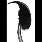
Transmission electron micrograph of longitudinal section through mouse spermatoz...
CIL:11831
NCBI Organism Classification
Cricetulus griseus
Biological Process
nucleus organization
Cellular Component
nucleus
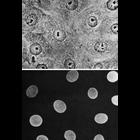
Light microscopy of the same field of cells showing the phase contrast image of ...