Alternate header for print version
Advanced search
Contributors
Help
Submit
Search
menu
Cell Process
Cell Component
Cell Type
Organism
Microbial
Alzheimer's
Data Sets
University of California, San Diego
9500 Gilman Drive
La Jolla, CA 92093-0608, USA
Voice
: (858) 534-0276
Fax
: (858) 534-7497
Email
: dorloff@ncmir.ucsd.edu
Grouped images - the images shown below are related
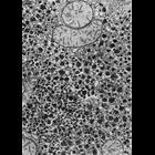
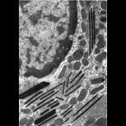
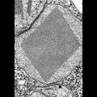
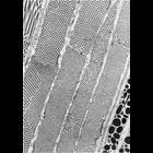
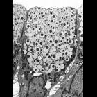
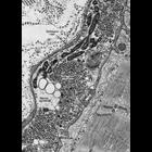
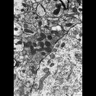
CIL:35972
NCBI Organism Classification
Phodopus
Biological Process
energy reserve metabolic process
Cellular Component
smooth endoplasmic reticulum
Figure 349 from Chapter 15 (Cytoplasmic Inclusions) of 'The Cell, 2nd Ed.' by Do...
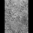
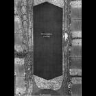
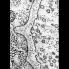
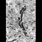
CIL:35973
NCBI Organism Classification
Notophthalmus
Biological Process
energy reserve metabolic process
Cellular Component
endoplasmic reticulum
Figure 350 from Chapter 15 (Cytoplasmic Inclusions) of 'The Cell, 2nd Ed.' by Do...
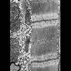
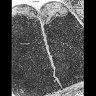
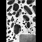
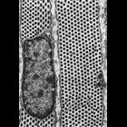
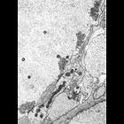
CIL:35974
NCBI Organism Classification
Felis catus
Biological Process
energy reserve metabolic process
Cellular Component
sarcoplasmic reticulum
Figure 351 from Chapter 15 (Cytoplasmic Inclusions) of 'The Cell, 2nd Ed.' by Do...
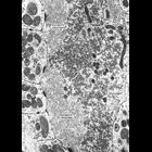
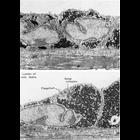
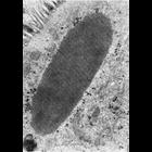
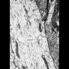
CIL:35975
NCBI Organism Classification
Chrysemys picta
Biological Process
energy reserve metabolic process
Cellular Component
synapse part
Figure 352 from Chapter 15 (Cytoplasmic Inclusions) of 'The Cell, 2nd Ed.' by Do...
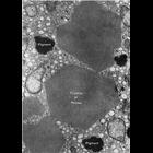
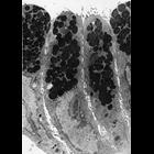
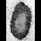
CIL:35976
NCBI Organism Classification
Cavia porcellus
Biological Process
energy reserve metabolic process
Cellular Component
glycogen granule
Figures 353 (upper) and 354 (lower) from Chapter 15 (Cytoplasmic Inclusions) of ...
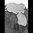
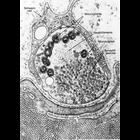
CIL:35977
NCBI Organism Classification
Cavia porcellus
Biological Process
energy reserve metabolic process
Cellular Component
glycogen granule
Figure 355 from Chapter 15 (Cytoplasmic Inclusions) of 'The Cell, 2nd Ed.' by Do...
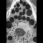
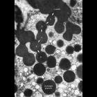
CIL:35978
NCBI Organism Classification
Didelphis virginiana
Biological Process
lipid storage
Cellular Component
lipid particle
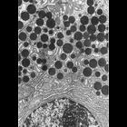
Figure 356 from Chapter 15 (Cytoplasmic Inclusions) of 'The Cell, 2nd Ed.' by Do...
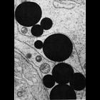
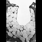
CIL:35979
NCBI Organism Classification
Rattus
Biological Process
lipid storage
Cellular Component
lipid particle
Figure 357 from Chapter 15 (Cytoplasmic Inclusions) of 'The Cell, 2nd Ed.' by Do...
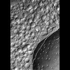
CIL:35980
NCBI Organism Classification
Litocranius walleri
Biological Process
lipid storage
Cellular Component
lipid particle
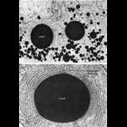
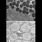
Figures 358 (upper) and 359 (lower) from Chapter 15 (Cytoplasmic Inclusions) of ...
CIL:35981
NCBI Organism Classification
Spermophilus citellus
Biological Process
lipid storage
Cellular Component
lipid particle
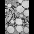
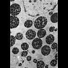
Figure 360 from Chapter 15 (Cytoplasmic Inclusions) of 'The Cell, 2nd Ed.' by Do...
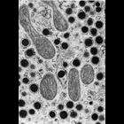
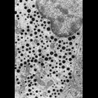
CIL:35983
NCBI Organism Classification
Rattus
Biological Process
lipid metabolic process
Cellular Component
lipid particle
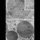
Figure 362 from Chapter 15 (Cytoplasmic Inclusions) of 'The Cell, 2nd Ed.' by Do...
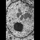
CIL:35984
NCBI Organism Classification
Batrachoseps attenuatus
Biological Process
none specified
Cellular Component
inclusion body
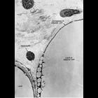
Figure 363 from Chapter 15 (Cytoplasmic Inclusions) of 'The Cell, 2nd Ed.' by Do...
CIL:35985
NCBI Organism Classification
Batrachoseps attenuatus
Biological Process
none specified
Cellular Component
inclusion body
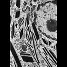
Figure 364 from Chapter 15 (Cytoplasmic Inclusions) of 'The Cell, 2nd Ed.' by Do...
CIL:35986
NCBI Organism Classification
Homo sapiens
Biological Process
none specified
Cellular Component
inclusion body
Figure 365 from Chapter 15 (Cytoplasmic Inclusions) of 'The Cell, 2nd Ed.' by Do...
CIL:35987
NCBI Organism Classification
Homo sapiens
Biological Process
none specified
Cellular Component
nuclear inclusion body
Figure 366 from Chapter 15 (Cytoplasmic Inclusions) of 'The Cell, 2nd Ed.' by Do...
CIL:35988
NCBI Organism Classification
Homo sapiens
Biological Process
none specified
Cellular Component
inclusion body
Figure 367 from Chapter 15 (Cytoplasmic Inclusions) of 'The Cell, 2nd Ed.' by Do...
CIL:35989
NCBI Organism Classification
Nicotiana tabacum
Biological Process
none specified
Cellular Component
microbody
Figure 368 from Chapter 15 (Cytoplasmic Inclusions) of 'The Cell, 2nd Ed.' by Do...
CIL:35990
NCBI Organism Classification
Felis catus
Biological Process
hemoglobin catabolic process
Cellular Component
inclusion body
Figure 369 from Chapter 15 (Cytoplasmic Inclusions) of 'The Cell, 2nd Ed.' by Do...
CIL:35991
NCBI Organism Classification
Rana pipiens
Biological Process
embryo development
Cellular Component
yolk
Figure 370 from Chapter 15 (Cytoplasmic Inclusions) of 'The Cell, 2nd Ed.' by Do...
CIL:35992
NCBI Organism Classification
Campylenchia latipes
Biological Process
none specified
Cellular Component
inclusion body
Figure 371 from Chapter 15 (Cytoplasmic Inclusions) of 'The Cell, 2nd Ed.' by Do...
CIL:35993
NCBI Organism Classification
Felis catus
Biological Process
detection of light stimulus involved in visual perception
Cellular Component
inclusion body
Figure 372 from Chapter 15 (Cytoplasmic Inclusions) of 'The Cell, 2nd Ed.' by Do...
CIL:35994
NCBI Organism Classification
Felis catus
Biological Process
detection of light stimulus involved in visual perception
Cellular Component
inclusion body
Figure 373 from Chapter 15 (Cytoplasmic Inclusions) of 'The Cell, 2nd Ed.' by Do...
CIL:35995
NCBI Organism Classification
Rhipicephalus appendiculatus
Biological Process
secretory granule organization
Cellular Component
secretory granule
Figure 374 from Chapter 15 (Cytoplasmic Inclusions) of 'The Cell, 2nd Ed.' by Do...
CIL:35996
NCBI Organism Classification
Rattus
Biological Process
secretory granule organization
Cellular Component
secretory granule
Figure 375 from Chapter 15 (Cytoplasmic Inclusions) of 'The Cell, 2nd Ed.' by Do...
CIL:35997
NCBI Organism Classification
Homo sapiens
Biological Process
secretory granule organization
Cellular Component
secretory granule
Figure 376 from Chapter 15 (Cytoplasmic Inclusions) of 'The Cell, 2nd Ed.' by Do...
CIL:35998
NCBI Organism Classification
Homo sapiens
Biological Process
secretory granule organization
Cellular Component
secretory granule
Figures 377 (upper) and 378 (lower) from Chapter 15 (Cytoplasmic Inclusions) of ...
CIL:35999
NCBI Organism Classification
Gerbillinae
Biological Process
secretory granule organization
Cellular Component
secretory granule
Figures 379 (upper) and 380 (lower) from Chapter 15 (Cytoplasmic Inclusions) of ...
CIL:36000
NCBI Organism Classification
Rhipicephalus appendiculatus
Biological Process
secretory granule organization
Cellular Component
secretory granule
Figure 381 from Chapter 15 (Cytoplasmic Inclusions) of 'The Cell, 2nd Ed.' by Do...
CIL:36001
NCBI Organism Classification
Rhipicephalus appendiculatus
Biological Process
secretory granule organization
Cellular Component
secretory granule
Figures 382 (upper) and 383 (lower) from Chapter 15 (Cytoplasmic Inclusions) of ...
CIL:36002
NCBI Organism Classification
Rattus
Biological Process
secretory granule organization
Cellular Component
secretory granule
Figure 384 from Chapter 15 (Cytoplasmic Inclusions) of 'The Cell, 2nd Ed.' by Do...
CIL:36003
NCBI Organism Classification
Cavia porcellus
Biological Process
secretory granule organization
Cellular Component
secretory granule
Figure 385 from Chapter 15 (Cytoplasmic Inclusions) of 'The Cell, 2nd Ed.' by Do...
CIL:36004
NCBI Organism Classification
none specified
Biological Process
exocytosis
Cellular Component
secretory granule
Figure 386 from Chapter 15 (Cytoplasmic Inclusions) of 'The Cell, 2nd Ed.' by Do...
CIL:36005
NCBI Organism Classification
Canis lupus familiaris
Biological Process
exocytosis
Cellular Component
secretory granule
Figure 387 from Chapter 15 (Cytoplasmic Inclusions) of 'The Cell, 2nd Ed.' by Do...
CIL:36006
NCBI Organism Classification
Leporidae
Biological Process
exocytosis
Cellular Component
secretory granule
Figure 388 from Chapter 15 (Cytoplasmic Inclusions) of 'The Cell, 2nd Ed.' by Do...
CIL:36007
NCBI Organism Classification
Homo sapiens
Biological Process
endocrine process
Cellular Component
secretory granule
Figure 389 from Chapter 15 (Cytoplasmic Inclusions) of 'The Cell, 2nd Ed.' by Do...
CIL:36008
NCBI Organism Classification
Homo sapiens
Biological Process
endocrine process
Cellular Component
secretory granule
Figure 390 from Chapter 15 (Cytoplasmic Inclusions) of 'The Cell, 2nd Ed.' by Do...
CIL:36009
NCBI Organism Classification
Rattus
Biological Process
secretory granule organization
Cellular Component
secretory granule
Figure 391 from Chapter 15 (Cytoplasmic Inclusions) of 'The Cell, 2nd Ed.' by Do...
CIL:36010
NCBI Organism Classification
Anura
Biological Process
synapse organization
Cellular Component
synapse
Figure 392 from Chapter 15 (Cytoplasmic Inclusions) of 'The Cell, 2nd Ed.' by Do...
CIL:36011
NCBI Organism Classification
Anura
Biological Process
synapse organization
Cellular Component
synapse
Figure 393 from Chapter 15 (Cytoplasmic Inclusions) of 'The Cell, 2nd Ed.' by Do...
CIL:36012
NCBI Organism Classification
Anura
Biological Process
synaptic vesicle exocytosis
Cellular Component
synapse
Figure 394 from Chapter 15 (Cytoplasmic Inclusions) of 'The Cell, 2nd Ed.' by Do...
CIL:36013
NCBI Organism Classification
Petromyzon marinus
Biological Process
synapse organization
Cellular Component
synapse
Figure 395 from Chapter 15 (Cytoplasmic Inclusions) of 'The Cell, 2nd Ed.' by Do...
CIL:36014
NCBI Organism Classification
Rattus
Biological Process
synapse organization
Cellular Component
synapse
Figure 396 from Chapter 15 (Cytoplasmic Inclusions) of 'The Cell, 2nd Ed.' by Do...
CIL:36015
NCBI Organism Classification
Rattus
Biological Process
synapse organization
Cellular Component
synapse
Figure 397 from Chapter 15 (Cytoplasmic Inclusions) of 'The Cell, 2nd Ed.' by Do...
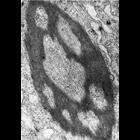
CIL:36016
NCBI Organism Classification
Rattus
Biological Process
synapse organization
Cellular Component
excitatory synapse
Figure 398 from Chapter 15 (Cytoplasmic Inclusions) of 'The Cell, 2nd Ed.' by Do...