Alternate header for print version


Licensing
Public Domain: This image is in the public domain and thus free of any copyright restrictions. However, as is the norm in scientific publishing and as a matter of courtesy, any user should credit the content provider for any public or private use of this image whenever possible. Learn more
tweet  
*CIL – Cell Image Library accession number. Please use this to reference an image.

CIL:10013*  Cite 
Description

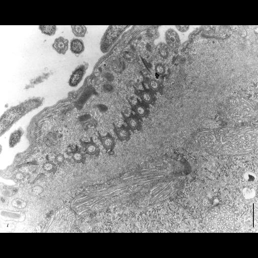
An oblique section shows the transverse aspect of basal bodies/cilia within one pectinelle of one of the two ciliary girdles of Didinium. Two ribbons of microtubules and a short kinetodesmal fiber arise from the proximal margin of each basal body. These may correspond to transverse microtubules and postciliary microtubules of other ciliates. Tips of parasomal sacs are also present. Microtubular ribbons under the alveoli seem to arise from extensions of the postciliary microtubules. TEM taken on 5/9/69 by R. Allen with Philips 300 operating at 60kV. Neg. 14,800X. Bar = 0.5µm. The negative was printed to paper and the image was scanned to Photoshop. This digitized image is available for qualitative analysis. A raw, unprocessed, high resolution version of this image (CIL:4663) is in the library and available for quantitative analysis. Standard glutaraldehyde fixation followed by osmium tetroxide, dehydrated in alcohol and embedded in an epoxy resin. Microtome sections prepared at approximately 75nm thickness. Additional information available at (http://www5.pbrc.hawaii.edu/allen/).

Biological Sources
NCBI Organism Classification
Didinium nasutum
Cell Type
eukaryotic cell
Eukaryotic Protist
Ciliated Protist
Cell Line
Carolina Biological Supply Company, NC, U.S.A.
Cellular Component
cell cortex
cilium
microtubule basal body
ciliary rootlet
bacterial ectosymbiont
Biological Context
Biological Process
ciliary cell motility
microtubule cytoskeleton organization
cytosketal organization
detection of symbiotic bacterium
Attribution
Names
Richard Allen
Link
http://www5.pbrc.hawaii.edu/allen/
Citation
Digital Object Identifier (DOI)
doi:10.7295/W9CIL10013
Archival Resource Key (ARK)
ark:/b7295/w9cil10013
Grouping This image is part of a group.
Imaging
Image Type
transmission electron microscopy (TEM)
illumination by electrons
Image Mode
detection of electrons
film
Parameters Imaged
electron density
Source of Contrast
stain with broad specificity
Visualization Methods
stain with broad specificity
osmium tetroxide
uranyl salt
lead salt
Processing History
film
Print from negative scanned for Photoshop.
Data Qualifiers
processed data
suitable for spatial measurements
Dimensions
Spatial Axis Image Size Pixel Size
X 3271px ——
Y 2634px ——