Alternate header for print version


Licensing
Attribution Non-Commercial; No Derivatives:This image is licensed under a Creative Commons Attribution, Non-Commercial, No Derivatives License. View License Deed | View Legal Code
tweet  
*CIL – Cell Image Library accession number. Please use this to reference an image.

CIL:10790*  Cite 
Description

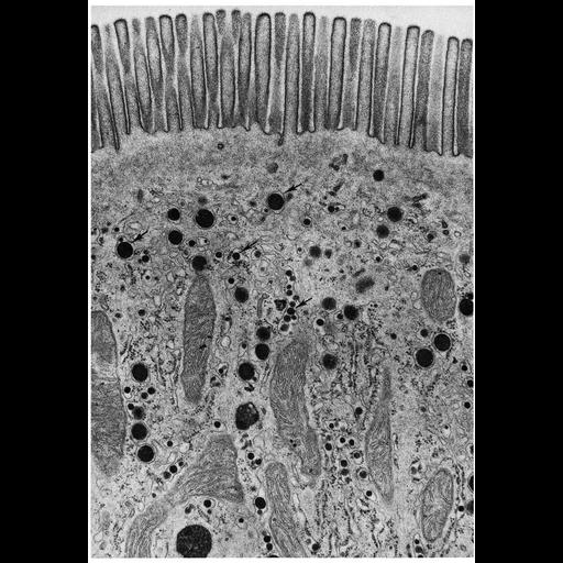
Figure 180 from Chapter 5 (Endoplasmic Reticulum) of 'The Cell, 2nd Ed.' by Don W. Fawcett M.D. Electron micrograph of a rat intestinal epithelium cell during absorption of a fatty meal. Droplets of lipid accumulate in the lumen of the smooth endoplasmic reticulum (arrows). Image by Sanford Palay. A PDF copy of the accompanying chapter is available on the ASCB’s BioEDUCATE website.

Biological Sources
NCBI Organism Classification
Rattus
Cell Type
brush border epithelial cell
Cellular Component
endoplasmic reticulum
smooth endoplasmic reticulum
Attribution
Names
Sanford Palay
Published
ISBN 0721635849
Link
http://www.ascb.org/bioeducate/FawcettTheCell...
Citation
Digital Object Identifier (DOI)
doi:10.7295/W9CIL10790
Archival Resource Key (ARK)
ark:/b7295/w9cil10790
Grouping This image is part of a group.
Imaging
Image Type
transmission electron microscopy (TEM)
Image Mode
illumination by electrons
detection of electrons
Parameters Imaged
electron density
Source of Contrast
stain with broad specificity
Data Qualifiers
processed data
Dimensions
Spatial Axis Image Size Pixel Size
X 894px ——
Y 1300px ——