Alternate header for print version


Licensing
Attribution Non-Commercial; No Derivatives:This image is licensed under a Creative Commons Attribution, Non-Commercial, No Derivatives License. View License Deed | View Legal Code
tweet  
*CIL – Cell Image Library accession number. Please use this to reference an image.

CIL:10817*  Cite 
Description

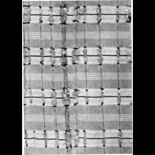
Figure 196 from Chapter 5 (Endoplasmic Reticulum) of 'The Cell, 2nd Ed.' by Don W. Fawcett M.D. A thin section through tibialis anterior of the mouse following fixation by glutaraldehyde solution lacking calcium, and postfixation with osmium-ferrocyanide solution. Skeletal tissue prepared this way results in staining of the etracellular space and T-tubules, but not the sacroplasmic reticulum. In this micrograph, two T-tubules can be seen in each sarcomere at the junctions of the A and I bands. Image by Michael Forbes. A PDF copy of the accompanying chapter is available on the ASCB’s BioEDUCATE website.

Biological Sources
NCBI Organism Classification
Mus musculus
Cell Type
skeletal muscle cell
Cellular Component
sarcoplasmic reticulum
myofibril
I band
A band
Z disc
terminal cisterna
sarcomere
Biological Context
Biological Process
striated muscle contraction
Attribution
Names
Michael Forbes
Published
ISBN 0721635849
Link
http://www.ascb.org/bioeducate/FawcettTheCell...
Citation
Digital Object Identifier (DOI)
doi:10.7295/W9CIL10817
Archival Resource Key (ARK)
ark:/b7295/w9cil10817
Grouping This image is part of a group.
Imaging
Image Type
transmission electron microscopy (TEM)
Image Mode
illumination by electrons
detection of electrons
Parameters Imaged
electron density
Source of Contrast
stain with broad specificity
osmium tetroxide
Data Qualifiers
processed data
Sample Preparation
Methods
glutaraldehyde fixed tissue
tissue in plastic embedment
Relation To Intact Cell
microtome-sectioned tissue
Dimensions
Spatial Axis Image Size Pixel Size
X 905px ——
Y 1276px ——