Alternate header for print version


Licensing
Attribution Non-Commercial; No Derivatives:This image is licensed under a Creative Commons Attribution, Non-Commercial, No Derivatives License. View License Deed | View Legal Code
tweet  
*CIL – Cell Image Library accession number. Please use this to reference an image.

CIL:10841*  Cite 
Description

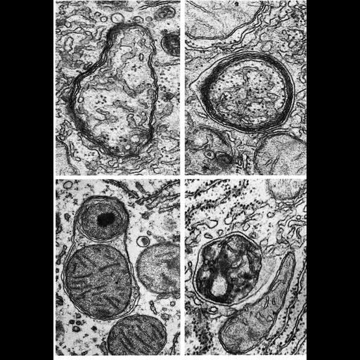
Figures 266 (upper left), 267 (upper right), 268 (lower left) and 269 (lower right) from Chapter 8 (Lysosomes) of 'The Cell, 2nd Ed.' by Don W. Fawcett M.D. Autophagic vacuoles from normal rat liver (Fig. 268, by Daniel Friend), and from liver several days after withdrawal of phenobarbital (Figs. 266, 267 and 269, by Robert Bolender). Content in the vacuoles in the upper panels is primarily smooth endoplasmic reticulum and free ribosomes. Vacuoles in the lower panels contain mitochondria and a peroxisome (lower left). A PDF copy of the accompanying chapter is available on the ASCB’s BioEDUCATE website.

Biological Sources
NCBI Organism Classification
Rattus
Cell Type
hepatocyte
Cellular Component
autophagic vacuole
autophagic vacuole membrane
endoplasmic reticulum
ribosome
mitochondrion
peroxisome
Biological Context
Biological Process
autophagy
Attribution
Names
Don W. Fawcett
Robert Bolender
Daniel Friend
Published
ISBN 0721635849
Link
http://www.ascb.org/bioeducate/FawcettTheCell...
Citation
Digital Object Identifier (DOI)
doi:10.7295/W9CIL10841
Archival Resource Key (ARK)
ark:/b7295/w9cil10841
Grouping This image is part of a group.
Imaging
Image Type
transmission electron microscopy (TEM)
Image Mode
illumination by electrons
detection of electrons
Parameters Imaged
electron density
Source of Contrast
stain with broad specificity
Data Qualifiers
processed data
Dimensions
Spatial Axis Image Size Pixel Size
X 898px ——
Y 1276px ——