Alternate header for print version


Licensing
Attribution Non-Commercial; No Derivatives:This image is licensed under a Creative Commons Attribution, Non-Commercial, No Derivatives License. View License Deed | View Legal Code
tweet  
*CIL – Cell Image Library accession number. Please use this to reference an image.

CIL:10929*  Cite 
Description

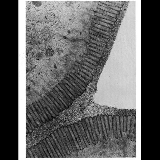
This electron micrograph shows a region of the brush border of the cat intestine. Tufted and branched polysaccharide filaments, each a few nanometers thick, extend from the microvilli to make up the glycocalyx, or surface coat, on the surface of plasma membrane of these epithelial cells. This image, courtesy of Susumu Ito is Figure 16 from Chapter 1 (The Cell Surface) of 'The Cell, 2nd Ed.' by Don W. Fawcett M.D. A PDF copy of the accompanying chapter is available on the ASCB's BioEDUCATE website.

Biological Sources
NCBI Organism Classification
Felis catus
Cell Type
epithelial cell
enterocyte
Cellular Component
plasma membrane
extracellular region part
glycocalyx
microvillus
Biological Context
Biological Process
extracellular structure organization
Attribution
Names
Susumu Ito
Published
ISBN 0721635849
Link
The Cell, 2nd Ed. by D.W. Fawcett
Citation
Digital Object Identifier (DOI)
doi:10.7295/W9CIL10929
Archival Resource Key (ARK)
ark:/b7295/w9cil10929
Grouping This image is part of a group.
Imaging
Image Type
transmission electron microscopy (TEM)
Image Mode
illumination by electrons
detection of electrons
Parameters Imaged
electron density
Source of Contrast
stain with broad specificity
Data Qualifiers
processed data
Sample Preparation
Methods
crosslinking-fixative fixed tissue
Relation To Intact Cell
microtome-sectioned tissue
Dimensions
Spatial Axis Image Size Pixel Size
X 996px ——
Y 1296px ——