Alternate header for print version


Licensing
Attribution Non-Commercial; No Derivatives:This image is licensed under a Creative Commons Attribution, Non-Commercial, No Derivatives License. View License Deed | View Legal Code
tweet  
*CIL – Cell Image Library accession number. Please use this to reference an image.

CIL:10946*  Cite 
Description

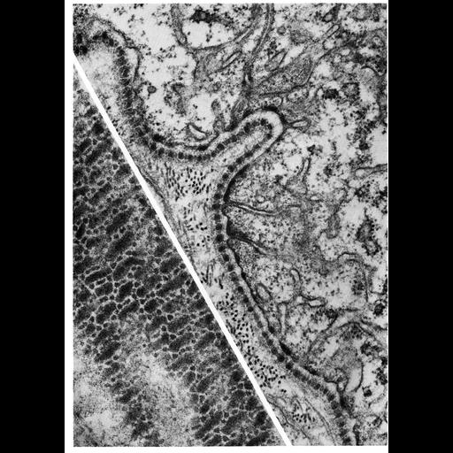
This electron micrograph shows the basal lamina along the midgut epithelium of the flea. Unlike the vertebrate basal lamina, which appears as a homogenous layer, the basal lamina in insects often appears striated. The inset panel offers a different view of the basal lamina in a section that is parallel to the base of the epithelium. In this view, the basal lamina is organized as a mosaic of elongated dense bodies with rows of smaller dense granules interspersed. Image by Susumu Ito, Figure 24 from Chapter 1 (The Cell Surface) of 'The Cell, 2nd Ed.' by Don W. Fawcett M.D. A PDF copy of the accompanying chapter is available on the ASCB's BioEDUCATE website.

Biological Sources
NCBI Organism Classification
Siphonaptera
Cell Type
epithelial cell
Cellular Component
basal lamina
extracellular region part
plasma membrane
Biological Context
Biological Process
extracellular structure organization
Attribution
Names
Susumu Ito
Published
ISBN 0721635849
Link
The Cell, 2nd Ed. by D.W. Fawcett
Citation
Digital Object Identifier (DOI)
doi:10.7295/W9CIL10946
Archival Resource Key (ARK)
ark:/b7295/w9cil10946
Grouping This image is part of a group.
Imaging
Image Type
transmission electron microscopy (TEM)
Image Mode
illumination by electrons
detection of electrons
Parameters Imaged
electron density
Source of Contrast
stain with broad specificity
Visualization Methods
electron-dense stain
Data Qualifiers
processed data
Sample Preparation
Methods
crosslinking-fixative fixed tissue
Relation To Intact Cell
microtome-sectioned tissue
Dimensions
Spatial Axis Image Size Pixel Size
X 910px ——
Y 1272px ——