Alternate header for print version


Licensing
Attribution Non-Commercial; No Derivatives:This image is licensed under a Creative Commons Attribution, Non-Commercial, No Derivatives License. View License Deed | View Legal Code
tweet  
*CIL – Cell Image Library accession number. Please use this to reference an image.

CIL:11014*  Cite 
Description

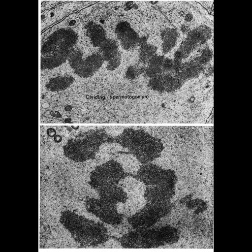
Transmission electron micrographs showing chromosomes in dividing spermatogonia during metaphase in tissue from ram testis (upper panel) and as they begin to separate during early anaphase in tissue from the Chinese hamster (lower panel).

Technical Details

Figures 124 (upper) and 125 (lower from Chapter 4 (Nucleus) of 'The Cell, 2nd Ed.' by Don W. Fawcett M.D. A PDF copy of the accompanying chapter is available on the ASCB's BioEDUCATE website.

Biological Sources
NCBI Organism Classification
Ovis aries
Cricetulus griseus
Cell Type
spermatogonium
Cellular Component
nuclear chromosome
Attribution
Names
Don W. Fawcett
Published
ISBN 0721635849
Published
from: Don W. Fawcett M.D ‘The Cell’ 2nd edition, W.B. Saunders Chapter 4 (Nucleus)
Link
ASCB BioEDUCATE Program
Citation
Digital Object Identifier (DOI)
doi:10.7295/W9CIL11014
Archival Resource Key (ARK)
ark:/b7295/w9cil11014
Grouping This image is part of a group.
Imaging
Image Type
film
Image Mode
transmission electron microscopy (TEM)
detection of electrons
Parameters Imaged
electron density
Source of Contrast
differences in adsorption or binding of stain
Visualization Methods
uranyl salt
osmium tetroxide
lead salt
Processing History
Image extracted from .pdf file of 'The Cell'
Data Qualifiers
processed data
Dimensions
Spatial Axis Image Size Pixel Size
X 900px ——
Y 1292px ——