Alternate header for print version


Licensing
Attribution Non-Commercial Share Alike:This image is licensed under a Creative Commons Attribution, Non-Commercial Share Alike License. View License Deed | View Legal Code
tweet  
*CIL – Cell Image Library accession number. Please use this to reference an image.

CIL:11041*  Cite 
Description

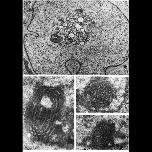
Transmission electron micrographs showing the unusual nucleolar structures in Sertoli cells of ruminants, with varying numbers of vesicles and tubules; the upper panel illustrates such an organization in the domestic bull, Bos taurus. Lower panels show the unusual nucleolar structures present in human endothelium, where a similar system of convoluted tubules is detected during the secretory phase of the menstrual cycle.

Technical Details

Upper panel is Figure 140; Lower panels appeared in Tersakis (1965) J. Cell Biol. 27: 158-61, reprinted with permission as Figures 141-3, from Chapter 4 (Nucleus) of 'The Cell, 2nd Ed.' by Don W. Fawcett M.D. A PDF copy of the corresponding chapter is available on the ASCB's BioEDUCATE website.

Biological Sources
NCBI Organism Classification
Barbus tauricus
Homo sapiens
Cell Type
Sertoli cell
glandular cell of endometrium
Cellular Component
nucleolus
nucleus
nuclear envelope
nuclear chromatin
Biological Context
Biological Process
nucleus organization
RNA metabolic process
nucleolus organization
Attribution
Names
Don W. Fawcett
John Tersakis
Published
ISBN 0721635849
Published
from: Don W. Fawcett M.D ‘The Cell’ 2nd edition, W.B. Saunders Chapter 4 (Nucleus)
Link
ASCB BioEDUCATE Program
Journal of Cell Biology
Citation
Digital Object Identifier (DOI)
doi:10.7295/W9CIL11041
Archival Resource Key (ARK)
ark:/b7295/w9cil11041
Grouping This image is part of a group.
Imaging
Image Type
film
Image Mode
transmission electron microscopy (TEM)
detection of electrons
Parameters Imaged
electron density
Source of Contrast
differences in adsorption or binding of stain
Visualization Methods
uranyl salt
osmium tetroxide
lead salt
Processing History
Image extracted from .pdf file of 'The Cell'
Data Qualifiers
processed data
Dimensions
Spatial Axis Image Size Pixel Size
X 891px ——
Y 1272px ——