Alternate header for print version


Licensing
Attribution Non-Commercial; No Derivatives:This image is licensed under a Creative Commons Attribution, Non-Commercial, No Derivatives License. View License Deed | View Legal Code
tweet  
*CIL – Cell Image Library accession number. Please use this to reference an image.

CIL:11046*  Cite 
Description

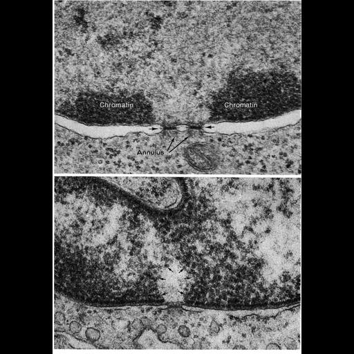
Transmission electron micrographs showing details of nuclear pores in thin sections of an erythroblast (above) and endothelial cell (below). Pores appear to be filled with dark staining material in these thin sections. Accumulations of heterochromatin at the nuclear envelope are excluded from pore-containing regions. Figures 146 and 147 from Chapter 4 (Nucleus) of 'The Cell' by Don W. Fawcett M.D.

Biological Sources
NCBI Organism Classification
Cavia sp.
Cell Type
erythroblast
Cellular Component
nuclear pore
nuclear envelope
nuclear chromatin
heterochromatin
Biological Context
Biological Process
nucleus organization
nuclear transport
nuclear pore organization
Attribution
Names
Don W. Fawcett
Published
ISBN 0721635849
Published
from: Don W. Fawcett M.D ‘The Cell’ 2nd edition, W.B. Saunders Chapter 4 (Nucleus)
Link
ASCB BioEDUCATE Program
Citation
Digital Object Identifier (DOI)
doi:10.7295/W9CIL11046
Archival Resource Key (ARK)
ark:/b7295/w9cil11046
Grouping This image is part of a group.
Imaging
Image Type
film
Image Mode
transmission electron microscopy (TEM)
detection of electrons
Parameters Imaged
electron density
Source of Contrast
differences in adsorption or binding of stain
Visualization Methods
uranyl salt
osmium tetroxide
lead salt
Processing History
Image extracted from .pdf file of 'The Cell'
Data Qualifiers
processed data
Dimensions
Spatial Axis Image Size Pixel Size
X 891px ——
Y 1268px ——