Alternate header for print version


Licensing
Attribution Non-Commercial; No Derivatives:This image is licensed under a Creative Commons Attribution, Non-Commercial, No Derivatives License. View License Deed | View Legal Code
tweet  
*CIL – Cell Image Library accession number. Please use this to reference an image.

CIL:11050*  Cite 
Description

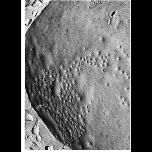
Transmission electron micrograph of surface of nuclear envelope of guinea pig spermatocyte prepared by the Freeze-fracture technique shows the unusual clustering of nuclear pores in these cells. From Fawcett and Chemes (1979) Tissue Cell 11:147-162, and reprinted with permission as Figure 150 from Chapter 4 (Nucleus) of 'The Cell, 2nd Ed.' by Don W. Fawcett M.D. A PDF copy of the accompanying chapter is available on the ASCB's BioEDUCATE website.

Technical Details

See: Fawcett and Chemes (1979) Tissue Cell 11:147-162

Biological Sources
NCBI Organism Classification
Cavia porcellus
Cell Type
spermatocyte
Cellular Component
nuclear envelope
nuclear pore
Biological Context
Biological Process
nucleus organization
nuclear transport
nuclear pore organization
Attribution
Names
Don W. Fawcett
Hector E. Chemes
Published
ISBN 0721635849
Published
from: Don W. Fawcett M.D ‘The Cell’ 2nd edition, W.B. Saunders Chapter 4 (Nucleus)
Pubmed
451991
Link
ASCB BioEDUCATE Program
Tissue and Cell
Citation
Digital Object Identifier (DOI)
doi:10.7295/W9CIL11050
Archival Resource Key (ARK)
ark:/b7295/w9cil11050
Grouping This image is part of a group.
Imaging
Image Type
film
Image Mode
transmission electron microscopy (TEM)
Parameters Imaged
electron density
Source of Contrast
differences in deposition of metal shadow
Visualization Methods
shadowing and plating
Processing History
Image extracted from .pdf file of 'The Cell'
Data Qualifiers
processed data
Sample Preparation
Methods
freeze_fracture/freeze_etch
Relation To Intact Cell
replica of fracture surface
Dimensions
Spatial Axis Image Size Pixel Size
X 914px ——
Y 1272px ——