Alternate header for print version


Licensing
Attribution Non-Commercial; No Derivatives:This image is licensed under a Creative Commons Attribution, Non-Commercial, No Derivatives License. View License Deed | View Legal Code
tweet  
*CIL – Cell Image Library accession number. Please use this to reference an image.

CIL:11106*  Cite 
Description

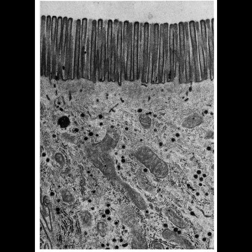
Electron micrograph showing the brush border of intestinal epithelial cells of the rat. The parallel microvilli of the brush border are about 0.1 µm wide and 1.0 µm in length. Image by Jean Paul Revel and Sanford Palay, Figure 38 from Chapter 2 (Specializations of the Free Surface) of 'The Cell, 2nd Ed.' by Don W. Fawcett M.D. A PDF copy of the accompanying chapter is available on the ASCB’s BioEDUCATE website.

Biological Sources
NCBI Organism Classification
Rattus
Cell Type
ciliated epithelial cell
Cellular Component
brush border
Biological Context
Biological Process
cell projection organization
Attribution
Names
Jean Paul Revel
Sanford Palay
Published
ISBN 0721635849
Link
Fawcett's "The Cell, 2nd Ed."
Citation
Digital Object Identifier (DOI)
doi:10.7295/W9CIL11106
Archival Resource Key (ARK)
ark:/b7295/w9cil11106
Grouping This image is part of a group.
Imaging
Image Type
transmission electron microscopy (TEM)
Image Mode
illumination by electrons
detection of electrons
Parameters Imaged
electron density
Source of Contrast
stain with broad specificity
Visualization Methods
osmium tetroxide
Data Qualifiers
processed data
Dimensions
Spatial Axis Image Size Pixel Size
X 926px ——
Y 1280px ——