Alternate header for print version


Licensing
Attribution Non-Commercial; No Derivatives:This image is licensed under a Creative Commons Attribution, Non-Commercial, No Derivatives License. View License Deed | View Legal Code
tweet  
*CIL – Cell Image Library accession number. Please use this to reference an image.

CIL:11129*  Cite 
Description

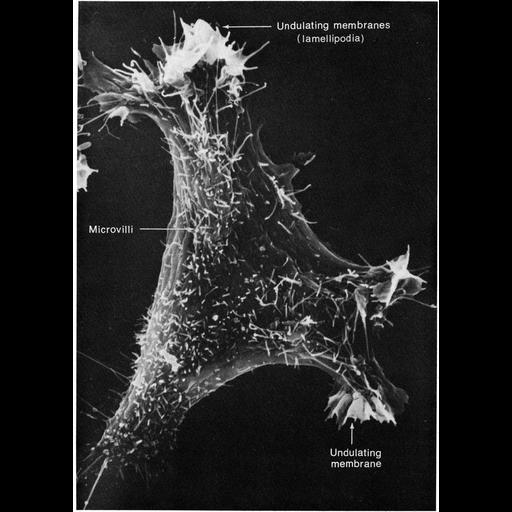
Scanning electron micrograph of a 3T3 cell growing in culture. Complexity and diversity of structure across the cell surface is apparent in the this image that s courtesy of Susan Brown. Figure 48 from Chapter 2 (Specializations of the Free Surface) of 'The Cell, 2nd Ed.' by Don W. Fawcett M.D. A PDF copy of the accompanying chapter is available on the ASCB’s BioEDUCATE website.

Biological Sources
NCBI Organism Classification
Mus musculus
Cell Type
fibroblast
Cell Line
3T3-Swiss albino
Cellular Component
lamellipodium membrane
microvillus
filopodium
cell surface
Biological Context
Biological Process
cell motility
cell projection organization
Attribution
Names
Susan Brown
Published
ISBN 0721635849
Link
Figure 47 from Chapter 2 (Specializations of ...
Citation
Digital Object Identifier (DOI)
doi:10.7295/W9CIL11129
Archival Resource Key (ARK)
ark:/b7295/w9cil11129
Grouping This image is part of a group.
Imaging
Image Type
scanning electron microscopy (SEM)
Image Mode
illumination by electrons
secondary_electron imaging
Parameters Imaged
secondary electron generation
Data Qualifiers
processed data
Sample Preparation
Methods
critical_point dried specimen
Relation To Intact Cell
dispersed cells in vitro
Dimensions
Spatial Axis Image Size Pixel Size
X 898px ——
Y 1256px ——