Alternate header for print version


Licensing
Attribution Non-Commercial; No Derivatives:This image is licensed under a Creative Commons Attribution, Non-Commercial, No Derivatives License. View License Deed | View Legal Code
tweet  
*CIL – Cell Image Library accession number. Please use this to reference an image.

CIL:11209*  Cite 
Description

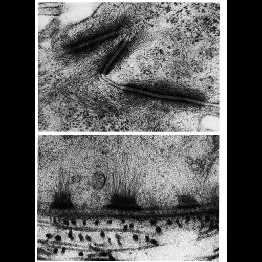
Upper panel, adjoining portions of two cells in the stratum spinosum of hamster cheek pouch epithelium shows desmosome junctions between two cells. Lower, a portion of the basal surface of a cell in the epidermis of a larval salamander Ambystoma punctatum shows hemi-desmosomes spaced at regular intervals. Figures 83 (upper, by J.T. Albright and M.A. Listgarten) and 84 (lower, by E. Hay) from Chapter 3 (Junctional Specializations) of 'The Cell, 2nd Ed.' by Don W. Fawcett M.D. A PDF copy of the accompanying chapter is available on the ASCB's BioEDUCATE website.

Biological Sources
NCBI Organism Classification
Phodopus
Ambystoma
Cell Type
squamous epithelial cell
Cellular Component
desmosome
cell-cell junction
keratin filament
cell-cell junction
Biological Context
Biological Process
cell adhesion
cell-cell junction organization
Attribution
Names
John Albright
M.A. Listgargen
Elizabeth Hay
Published
ISBN 0721635849
Link
The Cell, 2nd Ed. by D.W. Fawcett
Citation
Digital Object Identifier (DOI)
doi:10.7295/W9CIL11209
Archival Resource Key (ARK)
ark:/b7295/w9cil11209
Grouping This image is part of a group.
Imaging
Image Type
transmission electron microscopy (TEM)
Image Mode
illumination by electrons
detection of electrons
Parameters Imaged
electron density
Source of Contrast
stain with broad specificity
Visualization Methods
osmium tetroxide
Data Qualifiers
processed data
Dimensions
Spatial Axis Image Size Pixel Size
X 938px ——
Y 1284px ——