Alternate header for print version


Licensing
Attribution Non-Commercial; No Derivatives:This image is licensed under a Creative Commons Attribution, Non-Commercial, No Derivatives License. View License Deed | View Legal Code
tweet  
*CIL – Cell Image Library accession number. Please use this to reference an image.

CIL:11211*  Cite 
Description

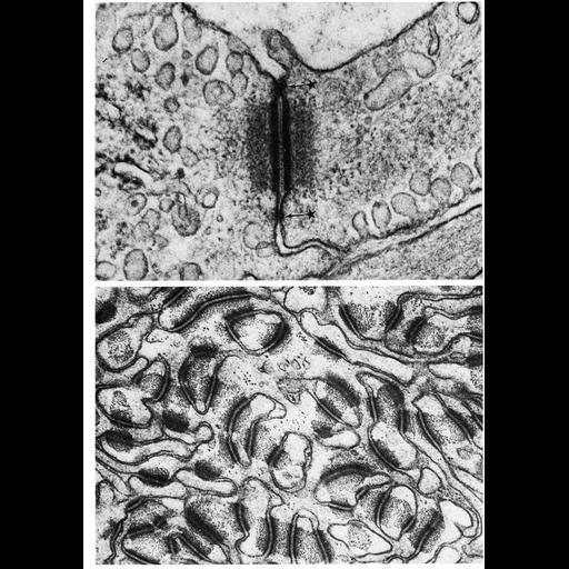
Upper: capillary endothelial cell junction in the rete mirabile of the gas bladder of the toadfish Opsanus tau shows dense intracelluular plaques and the central dense line in the intercellular space, characteristics that define desmosomes. Flanking this junction are other smaller and less highly organized densities with few, or no tonofilaments (asterisks). Lower: Desmosomes are abundant in the stratum spinosum of the epidermis from the muzzle of the cow, Bos taurus, an epithelium that is constantly subjected to flexion and attrition during grazing. Lower image contributed by Gida Matoltsy. Figures 85 (upper) and 86 (lower) from Chapter 3 (Junctional Specializations) of 'The Cell, 2nd Ed.' by Don W. Fawcett M.D. A PDF copy of the accompanying chapter is available on the ASCB's BioEDUCATE website.

Biological Context
Biological Process
cell adhesion
cell-cell junction organization
Attribution
Names
Don W. Fawcett
Gida Matoltsy
Published
ISBN 0721635849
Link
The Cell, 2nd Ed. by D.W. Fawcett
Citation
Digital Object Identifier (DOI)
doi:10.7295/W9CIL11211
Archival Resource Key (ARK)
ark:/b7295/w9cil11211
Grouping This image is part of a group.
Imaging
Image Type
transmission electron microscopy (TEM)
Image Mode
illumination by electrons
detection of electrons
Parameters Imaged
electron density
Source of Contrast
stain with broad specificity
Visualization Methods
osmium tetroxide
Data Qualifiers
processed data
Dimensions
Spatial Axis Image Size Pixel Size
X 910px ——
Y 1292px ——