Alternate header for print version


Licensing
Attribution Non-Commercial; No Derivatives:This image is licensed under a Creative Commons Attribution, Non-Commercial, No Derivatives License. View License Deed | View Legal Code
tweet  
*CIL – Cell Image Library accession number. Please use this to reference an image.

CIL:11226*  Cite 
Description

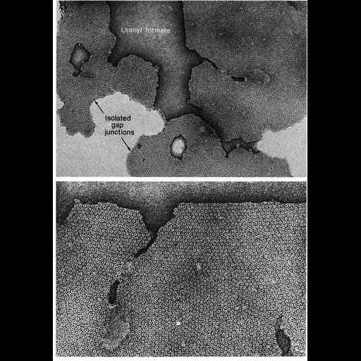
Gap junctions isolated from mouse liver and negatively stained with uranyl formate. At higher magnification, the 8-9 nm particles, or connexons, have a central 1.5-2 nm electron dense pore. Images by Bernard Gilula appear as Figures 97 (upper) and 98 (lower) from Chapter 3 (Junctional Specializations) of 'The Cell, 2nd Ed.' by Don W. Fawcett M.D. A PDF copy of the accompanying chapter is available on the ASCB's BioEDUCATE website.

Biological Sources
NCBI Organism Classification
Mus musculus
Cell Type
hepatocyte
Cellular Component
gap junction
connexon complex
Biological Context
Biological Process
cell communication
cell-cell junction organization
Attribution
Names
Bernard Gilula
Published
ISBN 0721635849
Link
The Cell, 2nd Ed. by D.W. Fawcett
Citation
Digital Object Identifier (DOI)
doi:10.7295/W9CIL11226
Archival Resource Key (ARK)
ark:/b7295/w9cil11226
Grouping This image is part of a group.
Imaging
Image Type
transmission electron microscopy (TEM)
Image Mode
illumination by electrons
detection of electrons
Parameters Imaged
electron density
Source of Contrast
stain with broad specificity
Visualization Methods
negative staining
Data Qualifiers
processed data
Sample Preparation
Relation To Intact Cell
isolated subcellular component
Dimensions
Spatial Axis Image Size Pixel Size
X 894px ——
Y 1268px ——