Alternate header for print version


Licensing
Attribution Non-Commercial; No Derivatives:This image is licensed under a Creative Commons Attribution, Non-Commercial, No Derivatives License. View License Deed | View Legal Code
tweet  
*CIL – Cell Image Library accession number. Please use this to reference an image.

CIL:11245*  Cite 
Description

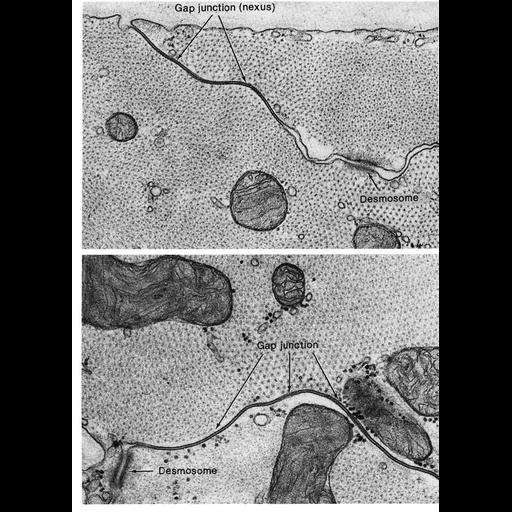
Gap junctions in papillary muscle from cat heart. These communicating junctions are responsible for the spreading wave of depolarization through the myocardium. Desmosomes can also be found on the lateral surfaces of cardiac muscle cells, often near gap junctions. Figures 107 (upper) and 108 (lower) from Chapter 3 (Junctional Specializations) of 'The Cell, 2nd Ed.' by Don W. Fawcett M.D. A PDF copy of the accompanying chapter is available on the ASCB's BioEDUCATE website.

Biological Sources
NCBI Organism Classification
Felis catus
Cell Type
cardiac muscle cell
Cellular Component
gap junction
desmosome
Biological Context
Biological Process
cell communication
cell-cell junction organization
Attribution
Names
Don W. Fawcett
Published
ISBN 0721635849
Link
The Cell, 2nd Ed. by D.W. Fawcett
Citation
Digital Object Identifier (DOI)
doi:10.7295/W9CIL11245
Archival Resource Key (ARK)
ark:/b7295/w9cil11245
Grouping This image is part of a group.
Imaging
Image Type
transmission electron microscopy (TEM)
Image Mode
illumination by electrons
detection of electrons
Parameters Imaged
electron density
Source of Contrast
stain with broad specificity
Visualization Methods
osmium tetroxide
Data Qualifiers
processed data
Dimensions
Spatial Axis Image Size Pixel Size
X 910px ——
Y 1268px ——