Alternate header for print version


Licensing
Attribution Non-Commercial; No Derivatives:This image is licensed under a Creative Commons Attribution, Non-Commercial, No Derivatives License. View License Deed | View Legal Code
tweet  
*CIL – Cell Image Library accession number. Please use this to reference an image.

CIL:11368*  Cite 
Description

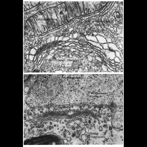
Figures 203 (upper panel, cell from Brunner's gland) and 204 (lower panel, alga) from Chapter 6 (Golgi Apparatus) of 'The Cell, 2nd Ed.' by Don W. Fawcett M.D. Transitional zone vesicles en route to the Golgi apparatus. In the upper panel, a ribosome-free region of the cistern of the endoplasmic reticulum is immediately below the mitochondrion, and numerous intermediate vesicles appear between the ER and the Golgi complex in a cell of Brunner's gland. Image by Daniel Friend. In the lower panel of an algal cell, vesicles can be seen budding from the perinuclear cistern. Lower panel from Massalski and Leedale (1969) Br. Phycol. J. 4:159-80, reprinted with permission. A PDF copy of the accompanying chapter is available on the ASCB’s BioEDUCATE website.

Biological Sources
Cell Type
secretory cell
Cellular Component
Golgi apparatus
Golgi stack
mitochondrion
endoplasmic reticulum
Attribution
Names
Daniel Friend
A. Massalski
G.F. Leedale
Published
ISBN 0721635849
Link
http://www.ascb.org/bioeducate/FawcettTheCell...
Citation
Digital Object Identifier (DOI)
doi:10.7295/W9CIL11368
Archival Resource Key (ARK)
ark:/b7295/w9cil11368
Grouping This image is part of a group.
Imaging
Image Type
transmission electron microscopy (TEM)
Image Mode
illumination by electrons
detection of electrons
Parameters Imaged
electron density
Source of Contrast
stain with broad specificity
Data Qualifiers
processed data
Sample Preparation
Methods
crosslinking-fixative fixed tissue
embedded tissue
Relation To Intact Cell
microtome-sectioned tissue
Dimensions
Spatial Axis Image Size Pixel Size
X 896px ——
Y 1304px ——