Alternate header for print version


Licensing
Attribution Non-Commercial; No Derivatives:This image is licensed under a Creative Commons Attribution, Non-Commercial, No Derivatives License. View License Deed | View Legal Code
tweet  
*CIL – Cell Image Library accession number. Please use this to reference an image.

CIL:11397*  Cite 
Description

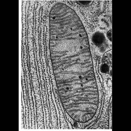
Figure 218 from Chapter 7 (Mitochondria) of 'The Cell, 2nd Ed.' by Don W. Fawcett M.D. The typical structural organization of mitochondria is revealed in this longitudinal section of a mitochondrion and surrounding cytoplasm from pancreas of the bat, Myotis lucifugus. It is bounded by a smooth outer membrane about 7nm thick. An inner membrane has infoldings called cristae that project into the interior of the organelle. Image by Keith Porter. A PDF copy of the accompanying chapter is available on the ASCB’s BioEDUCATE website.

Biological Context
Biological Process
cellular respiration
Attribution
Names
Keith Porter
Published
ISBN 0721635849
Link
Fawcett's "The Cell" 2nd Ed.
Citation
Digital Object Identifier (DOI)
doi:10.7295/W9CIL11397
Archival Resource Key (ARK)
ark:/b7295/w9cil11397
Grouping This image is part of a group.
Imaging
Image Type
transmission electron microscopy (TEM)
Image Mode
illumination by electrons
detection of electrons
Parameters Imaged
electron density
Source of Contrast
stain with broad specificity
Data Qualifiers
processed data
Dimensions
Spatial Axis Image Size Pixel Size
X 914px ——
Y 1280px ——