Alternate header for print version


Licensing
Attribution Non-Commercial; No Derivatives:This image is licensed under a Creative Commons Attribution, Non-Commercial, No Derivatives License. View License Deed | View Legal Code
tweet  
*CIL – Cell Image Library accession number. Please use this to reference an image.

CIL:11447*  Cite 
Description

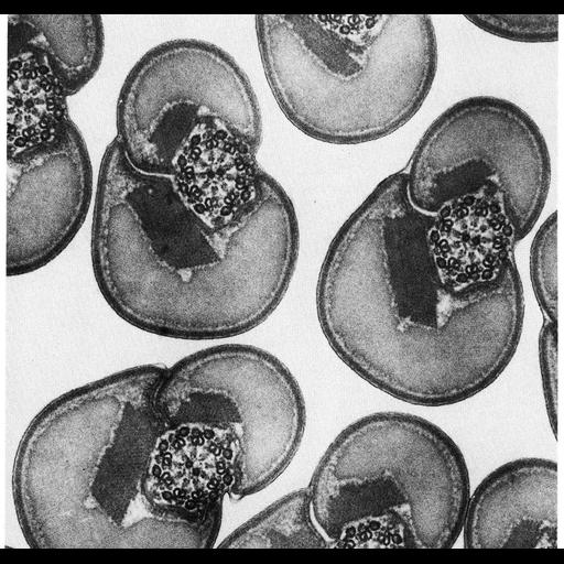
In insect spermiogenesis, mitochondrial cristae disappear and a proteinaceous material is deposited in filaments that become closely packed paracrystalline arrays. In this micrograph, crystals are present in each of the two mitochondrial derivatives that flank the axoneme in spermatozoa of the stilt bug, Jalysus. This image, courtesy of David Phillips, appears as Figure 252 from Chapter 7 (Mitochondria) of 'The Cell, 2nd Ed.' by Don W. Fawcett M.D. A PDF copy of the accompanying chapter is available on the ASCB’s BioEDUCATE website.

Biological Sources
NCBI Organism Classification
Jalysus
Cell Type
spermatid
Cellular Component
mitochondrial crista
axoneme
Biological Context
Biological Process
spermatogenesis
spermatid development
Attribution
Names
David Phillips
Published
ISBN 0721635849
Link
Fawcett's "The Cell" 2nd Ed.
Citation
Digital Object Identifier (DOI)
doi:10.7295/W9CIL11447
Archival Resource Key (ARK)
ark:/b7295/w9cil11447
Grouping This image is part of a group.
Imaging
Image Type
transmission electron microscopy (TEM)
Image Mode
illumination by electrons
detection of electrons
Parameters Imaged
electron density
Source of Contrast
stain with broad specificity
Data Qualifiers
processed data
Sample Preparation
Methods
crosslinking-fixative fixed tissue
embedded tissue
Relation To Intact Cell
microtome-sectioned tissue
Dimensions
Spatial Axis Image Size Pixel Size
X 906px ——
Y 859px ——