Alternate header for print version


Licensing
This image is copyright protected. Any public or private use of this image is subject to prevailing copyright laws. Please contact the content provider of this image for permission requests.
tweet  
*CIL – Cell Image Library accession number. Please use this to reference an image.

CIL:11556*  Cite 
Description

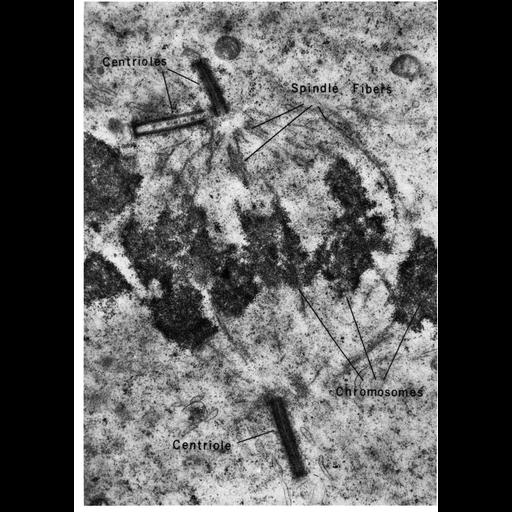
Figure 302 from Chapter 12 (Centrioles) by Don Fawcett. The centrioles replicate early in cell division and take up positions at either pole of the division figure. Concurrently with the condensation of the chromosomes and breakdown of the nuclear envelope, microtubules polymerize, extending from the chromosomes to the poles and from pole to pole, to form the mitotic spindle. In the accompanying micrograph made before introduction of aldehyde fixatives, the microtubules have not been well preserved. The plane of section is a fortunate one, however, in that three of the centrioles are included. The second centriole at the lower pole is in a plane perpendicular to this section and is not seen. Because the two diplosomes usually differ in their orientation, all four centrioles are rarely, if ever, included in the same thin section. A copy of the chapter is available on the ASCB's BioEDUCATE website.

Biological Sources
NCBI Organism Classification
Gallus gallus
Cell Type
spermatocyte
Cellular Component
pericentriolar material
nuclear chromosome
centriole
Biological Context
Biological Process
spermatogonial cell division
Attribution
Names
Don W. Fawcett
Toshio Nagano
Published
ISBN 0721635849
Link
http://www.ascb.org/bioeducate/FawcettTheCell...
Citation
Digital Object Identifier (DOI)
doi:10.7295/W9CIL11556
Archival Resource Key (ARK)
ark:/b7295/w9cil11556
Grouping This image is part of a group.
Imaging
Image Type
transmission electron microscopy (TEM)
illumination by electrons
Image Mode
detection of electrons
film
Parameters Imaged
electron density
Source of Contrast
stain with broad specificity
Visualization Methods
stain with broad specificity
osmium tetroxide
uranyl salt
lead salt
Processing History
recorded image
film
Print from negative scanned for Photoshop.
Data Qualifiers
processed data
Dimensions
Spatial Axis Image Size Pixel Size
X 920px ——
Y 1292px ——