Alternate header for print version


Licensing
Public Domain: This image is in the public domain and thus free of any copyright restrictions. However, as is the norm in scientific publishing and as a matter of courtesy, any user should credit the content provider for any public or private use of this image whenever possible. Learn more
tweet  
*CIL – Cell Image Library accession number. Please use this to reference an image.

CIL:122*  Cite 
Description

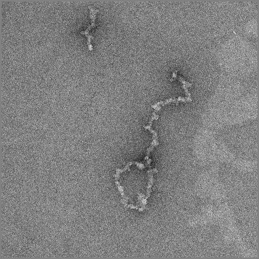
Telomeres isolated from chicken erythrocyte nuclei following restriction endonuclease treatment, and imaged by transmission EM after uranyl acetate negative staining. Sample was also labeled for the telomere protein TRF1 using immunogold. Image recorded with FEI Tecnai 12 TEM at 100KV and recorded on 2048x2048 CCD with 2x binning. Telomeres have a looped conformation, which likely aids in their protection from the rapid degradation that occurs with free DNA ends.

Biological Sources
NCBI Organism Classification
Gallus gallus gallus
Cell Type
erythrocyte
Cellular Component
chromosome, telomeric region
Biological Context
Biological Process
chromatin assembly
Molecular Function
structural constituent of chromatin
Attribution
Names
Christopher Woodcock
Citation
Digital Object Identifier (DOI)
doi:10.7295/W9CIL122
Archival Resource Key (ARK)
ark:/b7295/w9cil122
Imaging
Image Type
transmission electron microscopy (TEM)
Image Mode
illumination by electrons
detection of electrons
charge coupled device (CCD)
Parameters Imaged
electron density
Source of Contrast
elastic scattering
Visualization Methods
uranyl salt
uranium compound
gold
Processing History
unprocessed raw image
Sample Preparation
Methods
glutaraldehyde fixed tissue
Relation To Intact Cell
isolated subcellular component
Dimensions
Spatial Axis Image Size Pixel Size
X 1024px 0.454nm
Y 1024px 0.454nm