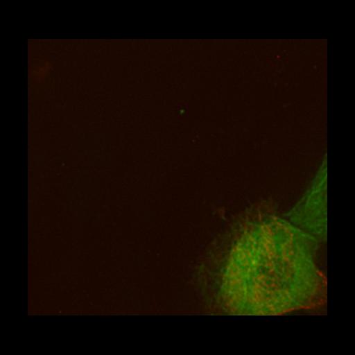
Behaviors of the microtubule anchoring factor RFP-LL5α (red) and microtubules (green) in living epithelial cells undergoing random migration. During the time-lapse imaging, the cells divided into two daughter cells. Cortical patches of RFP-LL5α appear when cells become attached to the glass substrate, but disappear when the cells become detached at the onset of cell division. The patches reappear soon after the cells adhere to the glass again. Overall findings from this publication showed that signaling from laminin-integrin associations plays a role in attaching microtubule plus ends to the epithelial basal cell cortex. This is the original data file for Video 4 from J Cell Biol (2010) 189 (5):901-917.
MCF-10Aeco cells expressing GFP-α-tubulin and RFP-LL5α were seeded on glass-bottomed dishes at a low density and cultured overnight. Time-lapse images of the cells were collected at 2.5-min intervals for 8 h 6 min using a DeltaVision Core Systemdriven by SoftWoRx software (Applied Precision) equipped with an IX70 microscope with a Plan-Apochromat 100× NA 1.40 oil immersion objective (Olympus), a cooled charge-coupled device camera (CoolSNAP HQ2; Photometrics) and a CO2 incubator (Tokai Hit Co., Ltd.). GFP, Ex, 470; Em, 525; RFP, Ex, 572, Em, 632.
| Spatial Axis | Image Size | Pixel Size |
|---|---|---|
| X | 432px | 0.127µm |
| Y | 399px | 0.127µm |
| Time | 150 seconds | 196 |
|---|