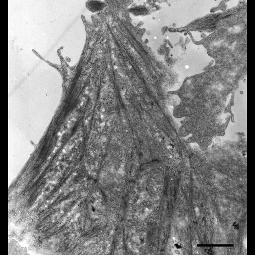
Network of stress fibers in an MDCK cell. This transmission electron micrograph of a flat-embedded MDCK cell was taken 60-70nm from the interface between the cell and the coverslip on which it was grown and fixed. The cells were cultured on Thermanox® coverslips, then chemically fixed with glutaraldehyde and osmium tetroxide before being stained en bloc with uranyl acetate. The cells were infiltrated with Spurr's® resin, placed under a vacuum for fifteen minutes, and then baked at 70° C. The coverslips were peeled away from the hardened resin wafers, which were then cut and thin-sectioned with a diamond knife. The 65 nm sections were placed on grids and post-stained with uranyl acetate and lead citrate. They were imaged using a Phillips CM 100 transmission electron microscope at an accelerating voltage of 80kV. Because the section was taken just inside the plasma membrane, many vesicles can be seen in this image, particularly in the lower left corner. A neighboring cell in the upper right is shown to underlap the main cell in this image. Scale bar = 2µm.
| Spatial Axis | Image Size | Pixel Size |
|---|---|---|
| X | 1706px | 0.0076µm |
| Y | 1818px | 0.0076µm |