Alternate header for print version


Licensing
This image is copyright protected. Any public or private use of this image is subject to prevailing copyright laws. Please contact the content provider of this image for permission requests.
tweet  
*CIL – Cell Image Library accession number. Please use this to reference an image.

CIL:12623*  Cite 
Description

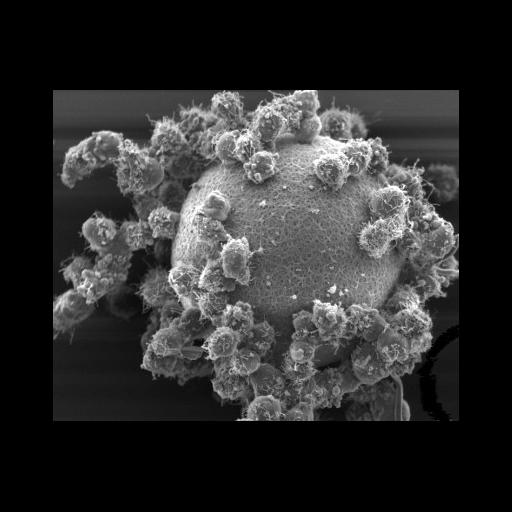
Hamster oocyte and cumulus cells. This scanning electron micrograph shows a hamster oocyte. The oocyte is not directly visible but lies beneath the zona pellucida. The surface of the zona pellucida can be seen between the cumulus cells that have remained attached to it. The sample was fixed using glutaraldehyde and osmium tetroxide, dehydrated in ethanol, critically point dried, coated with gold, and examined in a Phillips XL30 FEG scanning electron microscope. Magnification – 790x.

Biological Sources
NCBI Organism Classification
Mesocricetus auratus
Cell Type
oocyte
cumulus cell
Cellular Component
zona pellucida
Biological Context
Biological Process
ovulation
Attribution
Names
Prue Talbot
Citation
Digital Object Identifier (DOI)
doi:10.7295/W9CIL12623
Archival Resource Key (ARK)
ark:/b7295/w9cil12623
Imaging
Image Type
recorded image
Image Mode
scanning electron microscopy (SEM)
Parameters Imaged
elastic scattering of electrons
Source of Contrast
differences in deposition of metal shadow
Visualization Methods
gold
Processing History
unprocessed raw data
Data Qualifiers
raw, unprocessed data
Sample Preparation
Methods
glutaraldehyde fixed tissue
osmium tetroxide fixed tissue
dehydrated in ethanol
critical_point dried specimen
Relation To Intact Cell
whole mounted tissue
Dimensions
Spatial Axis Image Size Pixel Size
X 406px ——
Y 331px ——