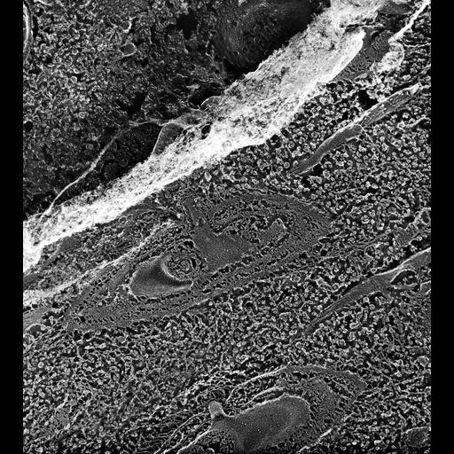
High resolution image of two parasomal sacs that are oriented perpendicular to the basal bodies. The top parasomal sac has a clathrin cage around it's coated pit area. The P-face of the plasma membrane has some IMPs, particularly around the neck of the basal body. Alveoli contain some non-etchable material. Striated bands are anchored to the cytosolic face of the inner alveolar membrane to which an inconspicuous epiplasm is bound. Fractured kinetodesmal fibers show evidence of longitudinally ordered packing of subunits even though cross striations are more prominent in thin sections. Additional interpretation is available at Chapter 8, Image 9 at the link below. TEM taken on 6/4/91 by R. Allen with Zeiss 10A operating at 80kV. Neg. 24,800X. The raw film was scanned with an Epson Perfection V750 Pro. This image is best used for quantitative analysis. Additional information available at (http://www5.pbrc.hawaii.edu/allen/).
| Spatial Axis | Image Size | Pixel Size |
|---|---|---|
| X | 3704px | 0.8nm |
| Y | 4120px | 0.8nm |