Alternate header for print version


Licensing
Public Domain: This image is in the public domain and thus free of any copyright restrictions. However, as is the norm in scientific publishing and as a matter of courtesy, any user should credit the content provider for any public or private use of this image whenever possible. Learn more
tweet  
*CIL – Cell Image Library accession number. Please use this to reference an image.

CIL:12639*  Cite 
Description

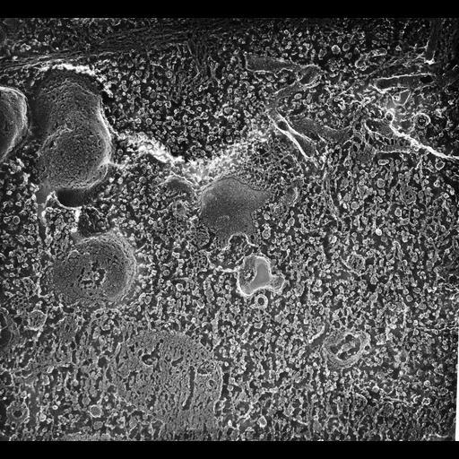
High resolution view of an early endosome with at least two coated evaginations showing, lies next to the proximal end of a fractured basal body. Other clathrin cages may indicate other evaginations from the same early endosome or from another nearby early endosome. TEM taken on 6/6/91 by R. Allen with Zeiss 10A operating at 80kV. Neg. 31,500X. Adapted with permission from J. Cell Sci. The raw film was scanned with an Epson Perfection V750 Pro. This image is best used for quantitative analysis. Additional information available at (http://www5.pbrc.hawaii.edu/allen/).

Biological Sources
NCBI Organism Classification
Paramecium multimicronucleatum
Cell Type
cell by organism
eukaryotic cell
Eukaryotic Protist
Ciliated Protist
Cellular Component
cytoplasm
cell cortex
endosome membrane
clathrin coat
microtubule basal body
Attribution
Names
Richard Allen (University of Hawaii)
Published
J. Cell Sci. 101:449-461, 1992
Pubmed
1629255
Link
http://www5.pbrc.hawaii.edu/allen/
Citation
Digital Object Identifier (DOI)
doi:10.7295/W9CIL12639
Archival Resource Key (ARK)
ark:/b7295/w9cil12639
Grouping This image is part of a group.
Dimensions
Spatial Axis Image Size Pixel Size
X 4310px 0.63nm
Y 3980px 0.63nm