Alternate header for print version


Licensing
Public Domain: This image is in the public domain and thus free of any copyright restrictions. However, as is the norm in scientific publishing and as a matter of courtesy, any user should credit the content provider for any public or private use of this image whenever possible. Learn more
tweet  
*CIL – Cell Image Library accession number. Please use this to reference an image.

CIL:12640*  Cite 
Description

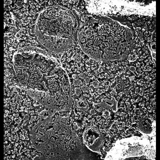
A high resolution image of a clump of at least seven small clathrin cages that are probably connected to an early endosome (below the fracture plane). The morphology of these cages shows a tighter packing than the larger clathrin cages at the parasomal sacs. TEM taken on 7/29/88 by C. Schroeder with Zeiss 10A operating at 80kV. Neg. 31,500X. The raw film was scanned with an Epson Perfection V750 Pro. This image is best used for quantitative analysis. Additional information available at (http://www5.pbrc.hawaii.edu/allen/).

Biological Sources
NCBI Organism Classification
Paramecium multimicronucleatum
Cell Type
cell by organism
eukaryotic cell
Eukaryotic Protist
Ciliated Protist
Cellular Component
cytoplasm
endosome
clathrin coat
Attribution
Names
Richard Allen (University of Hawaii)
C. Schroeder
Link
http://www5.pbrc.hawaii.edu/allen/
Citation
Digital Object Identifier (DOI)
doi:10.7295/W9CIL12640
Archival Resource Key (ARK)
ark:/b7295/w9cil12640
Grouping This image is part of a group.
Dimensions
Spatial Axis Image Size Pixel Size
X 3690px 0.63nm
Y 3878px 0.63nm