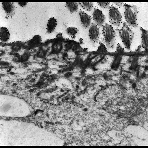
High resolution image of a monoclonal antibody raised in mouse to an antigen called C6 found on the surface of Paramecium. As seen in this micrograph the gold labelled antigen heavily labels the ciliary membrane and cell surface. In addition the gold is found in the parasomal sacs, in preendosomal vesicles and in early endosomes. However, there is almost no gold in the small coated evaginations of the early endosome or in the carrier vesicles escaping from the early endosomes. We conclude that this antigen passes no further into the cell and is recycled back to the plasma membrane. This antigen could be a cargo receptor that is cycled between the plasma membrane and the early endosome. The cargo may fall off this receptor in the early endosome and be concentrated in the small coated evaginations as is HRP for transport deeper into the cell. TEM taken on 5/14/91 by R. Allen with Zeiss 10A operating at 80kV. Neg. 12,000X. Adapted with permission from J. Cell Sci. 101:449-461, 1992. Cells were lightly fixed with 0.25% glutaraldehyde and infiltrated with 2.3M sucrose before being frozen in liquid nitrogen and thin sectioned at a temperature of –100°C at approximately 75nm thickness. Frozen sections from these preparations were then thawed, washed, and exposed to a monoclonal primary antibody that was raised in mice or rabbit/goat and to colloidal gold-complexed goat-anti-mouse/rabbit secondary antibodies. Further details of preparation are outlined in Methods Cell Biol. 2010;96:143-73. The raw film was scanned with an Epson Perfection V750 Pro. This image is best used for quantitative analysis. Additional information available at (http://www5.pbrc.hawaii.edu/allen/).
| Spatial Axis | Image Size | Pixel Size |
|---|---|---|
| X | 4862px | 1.25nm |
| Y | 4714px | 1.25nm |