Alternate header for print version


Licensing
Public Domain: This image is in the public domain and thus free of any copyright restrictions. However, as is the norm in scientific publishing and as a matter of courtesy, any user should credit the content provider for any public or private use of this image whenever possible. Learn more
tweet  
*CIL – Cell Image Library accession number. Please use this to reference an image.

CIL:1315*  Cite 
Description

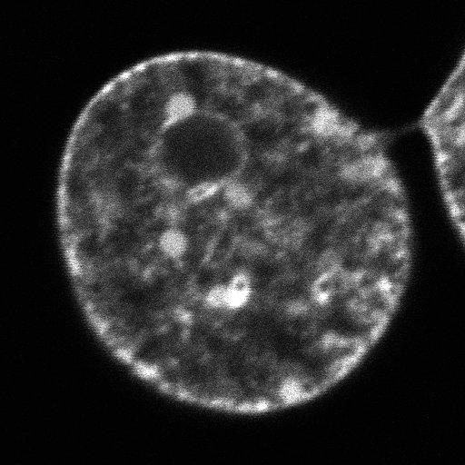
The intracellular mobility of MeCP2 (a methylated DNA-binding protein) followed in a mouse fibroblast stably expressing GFP-MeCP2 through a time series of images captured by confocal microscopy after bleaching a 2 micrometer circular area of euchromatin (center, right). Fluorescence recovery after photobleaching (FRAP) is rapid, indicating that MeCP2 in euchromatin is highly mobile.

Technical Details

Images were obtained using a Zeiss 510 Meta confocal microscope with excitation at 488 nm and emission between 505 nm and 530 nm. An initial scan of the cell was followed by the bleaching of a 2 um diameter spot of euchromatin, and 60 successive images recorded at 5 s intervals to measure fluorescence recovery due to MeCP2 mobility.

Biological Sources
NCBI Organism Classification
Mus musculus
Cell Type
fibroblast
Cellular Component
nucleus
euchromatin
nuclear chromatin
Biological Context
Biological Process
nucleus organization
chromatin organization
Attribution
Names
rajarshi ghosh
,/chris woodcock
Citation
Digital Object Identifier (DOI)
doi:10.7295/W9CIL1315
Archival Resource Key (ARK)
ark:/b7295/w9cil1315
Imaging
Image Type
recorded image
Image Mode
single-spot confocal microscopy
Parameters Imaged
fluorescence emission
Source of Contrast
fluorescence emission
Visualization Methods
fluorescent proteins derived from Aequorea victoria
Processing History
unprocessed raw data
Sample Preparation
Methods
whole mounted tissue
Relation To Intact Cell
living tissue
Dimensions
Spatial Axis Image Size Pixel Size
X 512px 0.04µm
Y 512px 0.04µm
Time 5 seconds 61