Alternate header for print version


Licensing
Attribution Non-Commercial Share Alike:This image is licensed under a Creative Commons Attribution, Non-Commercial Share Alike License. View License Deed | View Legal Code
tweet  
*CIL – Cell Image Library accession number. Please use this to reference an image.

CIL:13375*  Cite 
Description

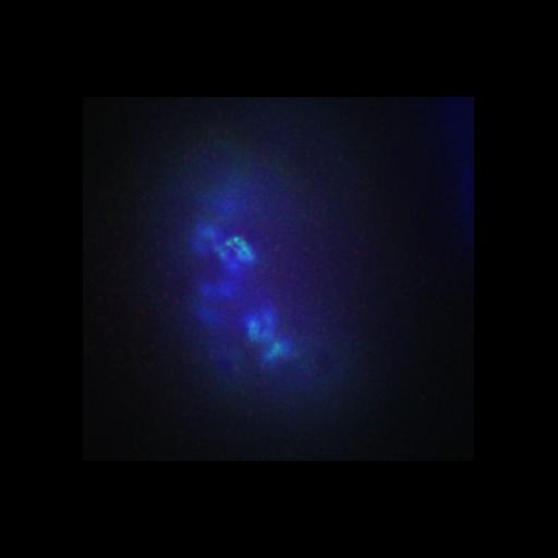
Z-focal series through a HeLa cell stained for DNA (blue), Aurora B (green), and phospho-Ser10-histone H3(red). This image is the control for the Bod-1 depletion experiment CIL: 13372. Bod1 is a protein that associates with a large macromolecular complex and localizes with kinetochores and spindle poles during mitosis.

Technical Details

Cells were fixed with 3.7% paraformaldehyde or methanol for 2 min at −20°C. Mouse anti–α-tubulin DM1A (Sigma-Aldrich), and mouse anti–Aurora B antibody AIM-1 (BD Biosciences) were used at 1:500. Fluorescently labeled secondary antibodies were all obtained from Jackson ImmunoResearch Laboratories. Image corresponds to Figure 5A from JCB vol. 179 no. 2 187-197.

Biological Sources
NCBI Organism Classification
Homo sapiens
Cell Line
HeLa S3
Cellular Component
chromosome, centromeric region
histone 3
spindle
DNA
Aurora B
cytoplasmic chromosome
Biological Context
Biological Process
mitosis
Aurora B activity
chromosome organization
Attribution
Names
Iain M. Porter
Sarah E. McClelland
Guennadi A. Khoudoli
Christopher J. Hunter
Jens S. Andersen
Andrew D. McAinsh
J. Julian Blow
Jason R. Swedlow
Published
JCB vol. 179 no. 2 187-197
Pubmed
17938248
Link
Journal of Cell Biology
Citation
Digital Object Identifier (DOI)
doi:10.7295/W9CIL13375
Archival Resource Key (ARK)
ark:/b7295/w9cil13375
Grouping This image is part of a group.
Sample Preparation
Methods
chemically fixed tissue
Relation To Intact Cell
dispersed cells in vitro
Dimensions
Spatial Axis Image Size Pixel Size
X 359px 0.0629µm
Y 334px 0.0629µm
Z 57px 0.2µm