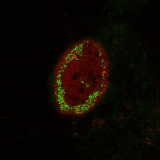
Upon mitochondrial uncoupling, transmembrane domain-deleted PINK1-YFP (green) fails to recruit mCherry-Parkin (red) to mitochondria in PINK1 knock-out mouse embryonic fibroblasts (PINK1 KO MEFs). Transfected PINK1 KO MEFs were treated with the mitochondrial depolarizing agent CCCP (carbonyl cyanide-m-chlorophenyl hydrazone) (10µM) for 3 hours. Live cell imaging was performed on an LSM510 Meta (Carl Zeiss, Inc) with a 63x 1.4 NA oil differential interference contrast Plan Apo objective. Image contrast and brightness were adjusted in the LSM image browser (Zeiss). This image corresponds to Supplemental Figure S1c, bottom right row of J Cell Biol, 191: 933-942, 2010. Images in Supplemental Figure S1c include CIL#s 13725, 13726, 13727, 13728.
| Spatial Axis | Image Size | Pixel Size |
|---|---|---|
| X | 512px | 0.1395µm |
| Y | 512px | 0.1395µm |