Alternate header for print version


Licensing
Attribution Non-Commercial Share Alike:This image is licensed under a Creative Commons Attribution, Non-Commercial Share Alike License. View License Deed | View Legal Code
tweet  
*CIL – Cell Image Library accession number. Please use this to reference an image.

CIL:13753*  Cite 
Description

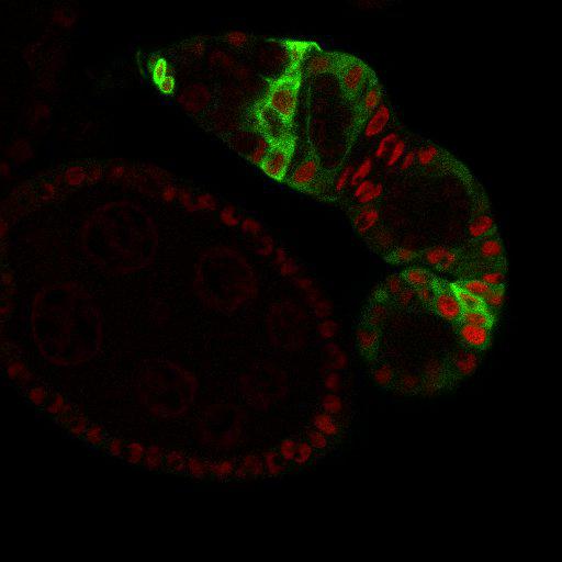
Expression of UAS-tauGFP (the tau-GFP fusion protein binds microtubules; green) under control of 109-30-Gal4 in wild-type Drosophila germaria. 109-30-Gal4 drives UAS-tauGFP expression in follicle stem cells and their progeny cells (see also CIL# 13752). Nuclei are in red. Image correlates to Supplemental Figure S2C in J Cell Biol. 2010. 191: 943-952.

Technical Details

Fly ovaries were dissected and fixed as described previously (O’Reilly et al., 2008). Primary antibody was anti-GFP. Secondary antibodies used were either FITC, Cy3, or Cy5 conjugated to species-specific secondary antibodies (Jackson ImmunoResearch Laboratories, Inc.). For nuclear staining, fixed ovaries were incubated for 15 min with Draq5 (Cell Signaling Technology). Samples were mounted in Vectashield mounting medium. Images were collected at room temperature (22C) using 40× (1.25 NA) or 63× (1.4 NA) oil immersion lenses (Leica) on an upright microscope (DM 5000; Leica) coupled to a confocal laser scanner (TCS SP5; Leica). LAS AF SP5 software (Leica) was used for data acquisition. Images representing individual channels of single confocal slices from the center of each germarium were exported as TIFF files, and images were converted to figures using Photoshop software (Adobe).

Biological Sources
NCBI Organism Classification
Drosophila melanogaster
Cell Type
germ line cell
follicle cell
germ line stem cell
follicle stem cell
Attribution
Names
Tiffiney R. Hartman
Daniel Zinshteyn
Heather K. Schofield
Emmanuelle Nicolas
Ami Okada
Alana M. O'Reilly
Published
J Cell Biol. 2010. 191: 943-952.
Pubmed
21098113
Link
http://jcb-dataviewer.rupress.org/jcb/browse/...
Citation
Digital Object Identifier (DOI)
doi:10.7295/W9CIL13753
Archival Resource Key (ARK)
ark:/b7295/w9cil13753
Grouping This image is part of a group.
Imaging
Image Type
recorded image
Image Mode
single-spot confocal microscopy
Parameters Imaged
fluorescence emission
Source of Contrast
distribution of a specific protein
compartmentalization of stain or label
Visualization Methods
primary antibody plus labeled secondary antibody
membrane-permeant probe
DRAQ5
EGFP
Processing History
unprocessed raw data
Data Qualifiers
raw, unprocessed data
suitable for spatial measurements
Sample Preparation
Methods
formaldehyde fixed tissue
detergent permeabilized
Relation To Intact Cell
whole mounted tissue
Dimensions
Spatial Axis Image Size Pixel Size
X 512px 0.2396µm
Y 512px 0.2396µm
Z 1px 0.2999µm