Alternate header for print version


Licensing
Attribution Non-Commercial Share Alike:This image is licensed under a Creative Commons Attribution, Non-Commercial Share Alike License. View License Deed | View Legal Code
tweet  
*CIL – Cell Image Library accession number. Please use this to reference an image.

CIL:150*  Cite 
Description

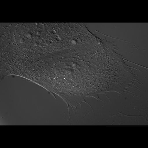
Differential interference microscopy (DIC) image of PtK1 cell (marsupial kidney epithelial cell expressing dominant-negative Rac1(T17N). Live cells were used to study microtubule dynamics using X-rhodamine tubulin (injected at 1 mg/mL)and EGFP-Rac1(T17N) (expression vector injected into the cell nucleus at 100 ug/mL). Image corresponds to Fig 3 and microtubule dynamics correspond to Video 3 in J Cell Biol, 161:845-851, 2003. Video 3 is contained within the same image group as this figure.

Biological Sources
NCBI Organism Classification
Potorous tridactylus
Cell Type
epithelial cell
Cell Line
Pt K1 (NBL-3)
Cellular Component
cell
nucleus
Biological Context
Molecular Function
dominant negative Rac
Attribution
Names
Torsten Wittmann
Gary M. Bokoch
Clare M. Waterman-Storer
Published
J Cell Biol, 161:845-851, 2003
Pubmed
12796474
Citation
Digital Object Identifier (DOI)
doi:10.7295/W9CIL150
Archival Resource Key (ARK)
ark:/b7295/w9cil150
Grouping This image is part of a group.
Imaging
Image Type
recorded image
Image Mode
differential interference contrast microscopy
Parameters Imaged
optical path length gradient
Source of Contrast
boundaries between regions with different refractive index
Visualization Methods
visualization of contiguous regions
Processing History
unprocessed raw data
Data Qualifiers
raw, unprocessed data
Sample Preparation
Methods
living tissue
Relation To Intact Cell
dispersed cells in vitro
Dimensions
Spatial Axis Image Size Pixel Size
X 1191px ——
Y 830px ——