Alternate header for print version


Licensing
Attribution Non-Commercial Share Alike:This image is licensed under a Creative Commons Attribution, Non-Commercial Share Alike License. View License Deed | View Legal Code
tweet  
*CIL – Cell Image Library accession number. Please use this to reference an image.

CIL:155*  Cite 
Description

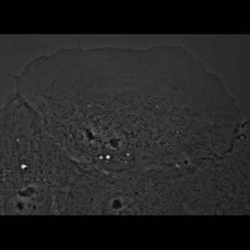
Phase contrast image of PtK1 cell (marsupial kidney epithelial cell) injected with Pak1 inhibitory fragment (H83L), X-rhodamine tubulin, and expressing constitutively active Rac(Q61L). Microtubule dynamics were visualized using X-rhodamine tubulin (injected at 1 mg/mL)and EGFP-Rac1(Q61L) (vector injected into nucleus). Corresponds to Fig 4B and Video 7 in J Cell Biol, 161:845-851, 2003. Video 7 is contained within the same image group as this figure.

Biological Sources
NCBI Organism Classification
Potorous tridactylus
Cell Type
epithelial cell
Cell Line
Pt K1 (NBL-3)
Cellular Component
cell
nucleus
lamellipodium
middle lamella
Biological Context
Molecular Function
Pak1 inhibition
active Rac
Attribution
Names
Torsten Wittmann
Gary M. Bokoch
Clare M. Waterman-Storer
Published
J Cell Biol, 161:845-851, 2003
Pubmed
12796474
Citation
Digital Object Identifier (DOI)
doi:10.7295/W9CIL155
Archival Resource Key (ARK)
ark:/b7295/w9cil155
Grouping This image is part of a group.
Imaging
Image Type
recorded image
Image Mode
phase contrast microscopy
Parameters Imaged
elastic scattering of photons
Source of Contrast
differences in amount of elastic light scattering
Visualization Methods
visualization of contiguous regions
Processing History
unprocessed raw data
Data Qualifiers
raw, unprocessed data
Sample Preparation
Methods
living tissue
Relation To Intact Cell
dispersed cells in vitro
Dimensions
Spatial Axis Image Size Pixel Size
X 1153px ——
Y 837px ——