Alternate header for print version


Licensing
Public Domain: This image is in the public domain and thus free of any copyright restrictions. However, as is the norm in scientific publishing and as a matter of courtesy, any user should credit the content provider for any public or private use of this image whenever possible. Learn more
tweet  
*CIL – Cell Image Library accession number. Please use this to reference an image.

CIL:21994*  Cite 
Description

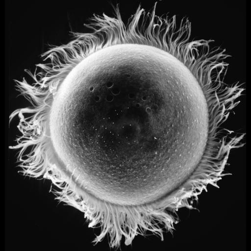
The aboral view of Didinium showing seven contractile vacuole pores for it's single posterior contractile vacuole and metachronous waves of cilia in one of the two characteristic ciliary girdles of Didinium nasutum. To one side you will note five white lines that represent rows of clavate cilia (also seen in CIL:17891 and CIL:19538). The bumps you will see on the surface at higher magnification may be either solitary clavate cilia or cyrtocysts, an extrusive organelle of unknown (possibly defensive) function. A thin section of a cyrtocyst can be seen at CIL:4665. This micrograph was taken in 1968 by G. Antipa on a Cambridge Mark IIA operating at 20kV. The negative magnification is 825X. The raw film was scanned with an Epson Perfection V750 Pro. This image is available for qualitative analysis. Further details are available at Wessenberg, H. and Antipa, G. 1968. Studies on Didinium nasutum. I. Structure and ultrastructure. Protistologica 4:427-447.

Biological Sources
NCBI Organism Classification
Didinium nasutum
Cell Type
cell by organism
eukaryotic cell
Eukaryotic Protist
Ciliated Protist
Cellular Component
contractile vacuole pore
cilium
cell cortex
cell surface
Attribution
Names
Gregory Antipa (San Francisco State University)
Citation
Digital Object Identifier (DOI)
doi:10.7295/W9CIL21994
Archival Resource Key (ARK)
ark:/b7295/w9cil21994
Grouping This image is part of a group.
Imaging
Image Type
recorded image
Image Mode
illumination by electrons
secondary_electron imaging
Parameters Imaged
illumination by electrons
secondary electron generation
secondary_electron imaging
Source of Contrast
detection of electrons
Visualization Methods
shadowing and plating
Processing History
recorded image
film
unprocessed raw data
Data Qualifiers
raw, unprocessed data
Sample Preparation
Methods
chemically fixed tissue
Parducz fixative
lyophilized specimen
Au/Pd metal sputter-coat
Relation To Intact Cell
whole mounted tissue
Dimensions
Spatial Axis Image Size Pixel Size
X 5437px 17.6nm
Y 5658px 17.6nm