Alternate header for print version


Licensing
Public Domain: This image is in the public domain and thus free of any copyright restrictions. However, as is the norm in scientific publishing and as a matter of courtesy, any user should credit the content provider for any public or private use of this image whenever possible. Learn more
tweet  
*CIL – Cell Image Library accession number. Please use this to reference an image.

CIL:22754*  Cite 
Description

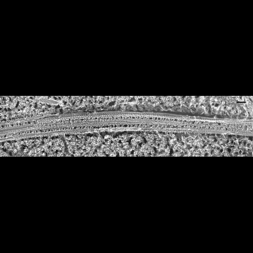
An isolated and longitudinally fractured cilium exposing the middle of the cilium. The spokes extend from the doublets toward the two singlet microtubules. These spokes are spaced unevenly so they appear to be arranged into triplets. The fracture plane, while in general is longitudinal, exposes the singlets and outer doublets at different levels so various periodicities appear along these microtubules. The cilium may also be slightly twisted along its length. TEM taken on 2/15/88 by C. Schroeder with Zeiss 10A operating at 80kV. Neg. 31,500X. Bar = 0.1µm. A print of the negative was scanned and processed in Photoshop. This image is best used for qualitative analysis. A high resolution image (CIL:12064) is available for quantitative analysis. Additional information available at (http://www5.pbrc.hawaii.edu/allen/).

Biological Sources
NCBI Organism Classification
Paramecium multimicronucleatum
Cell Type
cell by organism
eukaryotic cell
Eukaryotic Protist
Ciliated Protist
Cellular Component
cilium
radial spoke
axonemal microtubule
cilium axoneme
Biological Context
Biological Process
cilium axoneme assembly
cilium movement
cilium assembly
Attribution
Names
Richard Allen (University of Hawaii)
C. Schroeder
Citation
Digital Object Identifier (DOI)
doi:10.7295/W9CIL22754
Archival Resource Key (ARK)
ark:/b7295/w9cil22754
Grouping This image is part of a group.
Imaging
Image Type
transmission electron microscopy (TEM)
illumination by electrons
Image Mode
electron density
Parameters Imaged
elastic scattering of electrons
electron density
elevation
Source of Contrast
differences in deposition of metal shadow
Visualization Methods
transmission electron microscopy (TEM)
Processing History
freeze_fracture/freeze_etch
shadowing and plating
recorded image
film
Print of negative scanned for Photoshop
Data Qualifiers
processed data
Dimensions
Spatial Axis Image Size Pixel Size
X 2400px ——
Y 580px ——