Alternate header for print version


Licensing
Attribution Only: This image is licensed under a Creative Commons Attribution License. View License Deed | View Legal Code
tweet  
*CIL – Cell Image Library accession number. Please use this to reference an image.

CIL:25112*  Cite 
Description

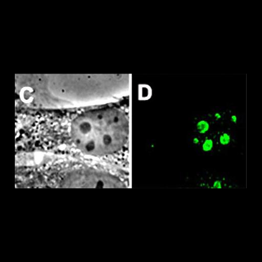
Mouse 3T3 fibroblast shown in phase contrast (left) and immuno-stained for the nucleolar protein nucleostemin (right, green).

Technical Details

Data were collected using a Leica DMIRB microscope equipped with a 100x objective (N.A. 1.4) and appropriate filter sets, and images captured using a Quantix 57 CCD camera (Roper Scientific Photometrics). For high resolution spatial mapping, three-dimensional optical stacks (containing 21 consecutive 0.25 micron slices) were captured using a PIFOC microscope focusing drive (Polytec PI). Images were dark current subtracted and intensity scaled See Fig 1 CD in Politz et al., 2005 Mol Biol Cell 16:3401-3410.

Biological Sources
NCBI Organism Classification
Mus musculus
Cell Type
fibroblast
Cell Line
3T3
Cellular Component
nucleolus
nucleus
Attribution
Names
Joan Politz
Published
Politz et al., 2005 Mol Biol Cell 16:3401-3410
Pubmed
15857956
Link
Molecular Biology of the Cell
OTHER
Ilvin Polena
Ian Trask
David Bazett-Jones
Thoru Pederson
Citation
Digital Object Identifier (DOI)
doi:10.7295/W9CIL25112
Archival Resource Key (ARK)
ark:/b7295/w9cil25112
Grouping This image is part of a group.
Imaging
Image Type
charge coupled device (CCD)
Image Mode
phase contrast microscopy
fluorescence microscopy
Parameters Imaged
elastic scattering of photons
fluorescence emission
Source of Contrast
differences in amount of elastic light scattering
distribution of epitope
Visualization Methods
Fluorescein (FITC)
Processing History
multipanel montage
Data Qualifiers
processed data
Sample Preparation
Methods
formaldehyde fixed tissue
Relation To Intact Cell
whole mounted tissue
Dimensions
Spatial Axis Image Size Pixel Size
X 453px ——
Y 224px ——