Alternate header for print version


Licensing
Attribution Non-Commercial Share Alike:This image is licensed under a Creative Commons Attribution, Non-Commercial Share Alike License. View License Deed | View Legal Code
tweet  
*CIL – Cell Image Library accession number. Please use this to reference an image.

CIL:26552*  Cite 
Description

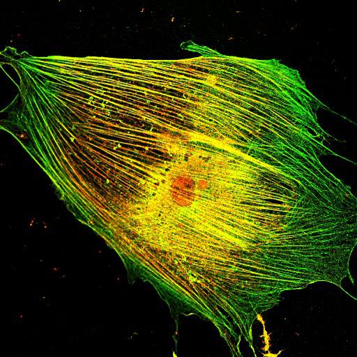
To study the molecular mechanism by which nonmuscle myosin II (MII) regulates protrusion and adhesion dynamics in migrating cells, Swiss3T3 cells were co-stained for TRIO (green) and myosin IIB (red). These findings help elucidate a functional link between MII and Rac1/Cdc42 GTPases, which may regulate protrusion/adhesion dynamics in migrating cells. This image is the original data file from Fig. 2F “colocalization of MII with GEFs”, J. Cell Biol. 2010. Vol. 190(4):663–674.

Technical Details

Cells were cultured in DME (Invitrogen) supplemented with 10% fetal bovine serum and 100 U/ml penicillin/streptomycin (Invitrogen) at 37°C in a humidified 5% CO2 incubator. Cells were serum-starved overnight, then fixed using 3.7% paraformaldehyde in PBS for 15 min, permeabilized using 0.2% Triton X-100 in PBS for 2 min, blocked with 2% BSA in PBS, and stained with the indicated primary antibodies at 4°C overnight, followed by incubation with a secondary Alexa Fluor 488–, 546– or 594–conjugated antibody. Images were captured by Leica TCS SP2 confocal microscope with HCX PL APO 63X objective.

Biological Sources
NCBI Organism Classification
Mus musculus
Cell Type
fibroblast
Cell Line
3T3-Swiss albino
Cellular Component
lamellipodium
myosin II complex
actin filament
Biological Context
Biological Process
regulation of cell migration
Attribution
Names
Chan-Soo Lee
Chang-Ki Choi
Eun-Young Shin
Martin Alexander Schwartz
Eung-Gook Kim
Published
J. Cell Biol. 2010. Vol. 190(4):663–674
Pubmed
PMID:20713598
Link
http://jcb-dataviewer.rupress.org/jcb/browse/...
Citation
Digital Object Identifier (DOI)
doi:10.7295/W9CIL26552
Archival Resource Key (ARK)
ark:/b7295/w9cil26552
Grouping This image is part of a group.
Imaging
Image Type
recorded image
Image Mode
confocal microscopy
Parameters Imaged
fluorescence emission
Source of Contrast
distribution of epitope
Visualization Methods
Alexa Fluor 488
Alexa Fluor 546
Data Qualifiers
raw, unprocessed data
Sample Preparation
Methods
formaldehyde fixed tissue
detergent permeabilized
Relation To Intact Cell
dispersed cells in vitro
Dimensions
Spatial Axis Image Size Pixel Size
X 512px ——
Y 512px ——