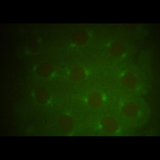
A confocal 4D-stack from Cycle 11 in a transgenic Drosophila embryo expressing gfp-tubulin (shown in green) and histone-rfp (shown in red). This image is original data contributing to Fig. S1. "Spindle dynamics in Drosophila embryos and in vitro gliding assays with Ncd and KLP61F" from Civelekoglu-Scholey et al.(2010) Prometaphase spindle maintenance by an antagonistic motor-dependent force balance made robust by a disassembling lamin-B envelope, J. Cell Biol. 188(1):49-68.
Embryos expressing gfp-tubulin and histone-rfp were collected at 25°C for 1 h, matured for 40 min, dechorionated, placed on heptane glue and covered with halocarbon oil. Images from this image group were acquired with an inverted IX-70 Olympus with Ultra-View spinning disk confcal head (PerkinElmer) and acquired with an oil immersion objective (UPlan-Apochromat 100x N.A. 1.35, or Plan-Apochromat 60x NA 1.4). Time series (at intervals of 3 to 10 sec) z-stacks of planes at 0.5 µm were acquired with an Orca II CCD camera (Hamamatsu Photonics). For further information see J. Cell Biol. 188(1):49-68.
| Spatial Axis | Image Size | Pixel Size |
|---|---|---|
| X | 672px | —— |
| Y | 512px | —— |
| Z | 4px | 0.5µm |