Alternate header for print version


Licensing
Attribution Non-Commercial Share Alike:This image is licensed under a Creative Commons Attribution, Non-Commercial Share Alike License. View License Deed | View Legal Code
tweet  
*CIL – Cell Image Library accession number. Please use this to reference an image.

CIL:34619*  Cite 
Description

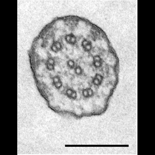
A cross section near the proximal end of the cilium. Triangular densities link the doublets to the membrane. Bar = 0.2µm. TEM taken 7/23/65 by R. Allen with RCA EMU3F operating at 50kV. Adapted with permission. The negative was printed to paper and the image was scanned to Photoshop. This digitized image is available for qualitative analysis. An unprocessed, high resolution version of this image (CIL:34604) is in the library and available for quantitative analysis. Additional information available at (http://www5.pbrc.hawaii.edu/allen/).

Technical Details

Standard glutaraldehyde fixation followed by osmium tetroxide, dehydrated in alcohol and embedded in an epoxy resin. Microtome sections prepared at approximately 75nm thickness. Additional information available at (http://www5.pbrc.hawaii.edu/allen/).

Biological Sources
NCBI Organism Classification
Tetrahymena pyriformis
Cell Type
cell by organism
eukaryotic cell
Eukaryotic Protist
Ciliated Protist
Cellular Component
cilium
cilium axoneme
Biological Context
Biological Process
cilium axoneme assembly
Attribution
Names
Richard Allen (University of Hawaii)
Citation
Digital Object Identifier (DOI)
doi:10.7295/W9CIL34619
Archival Resource Key (ARK)
ark:/b7295/w9cil34619
Grouping This image is part of a group.
Imaging
Image Type
transmission electron microscopy (TEM)
illumination by electrons
Image Mode
detection of electrons
film
Parameters Imaged
electron density
Source of Contrast
intrinsic mass distribution
stain with broad specificity
Visualization Methods
stain with broad specificity
uranyl salt
gold
Processing History
recorded image
film
Print from negative scanned for Photoshop.
Data Qualifiers
processed data
Sample Preparation
Methods
glutaraldehyde fixed tissue
tissue in vitreous ice embedment
microtome-sectioned tissue
monoclonal antibody
Relation To Intact Cell
microtome-sectioned tissue
Dimensions
Spatial Axis Image Size Pixel Size
X 586px ——
Y 756px ——