Alternate header for print version


Licensing
Attribution Non-Commercial Share Alike:This image is licensed under a Creative Commons Attribution, Non-Commercial Share Alike License. View License Deed | View Legal Code
tweet  
*CIL – Cell Image Library accession number. Please use this to reference an image.

CIL:34883*  Cite 
Description

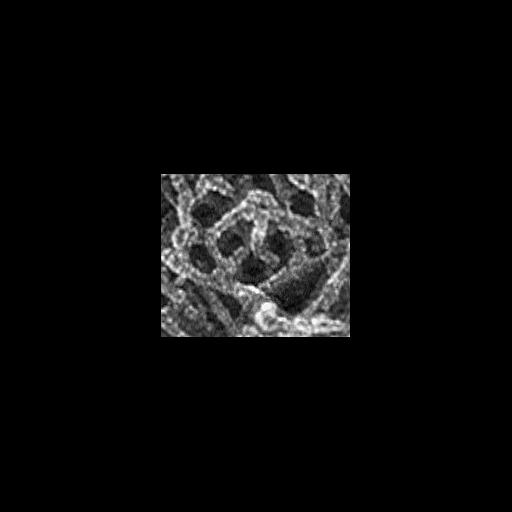
Multiple branching of actin filaments in lamellipodia of Xenopus keratocytes. This image show an enlargement of a local region from the overview of the leading edge, CIL 24786. Image corresponds to Figure 1c from J Cell Biol. 1999 May 31;145(5):1009-26. Figure 1b-1g are CIL 24882-24887.

Technical Details

Procedures for detergent extraction, immunostaining, S1 decoration, light, and EM were described previously (Svitkina et al., 1995, 1996, 1997;Verkhovsky et al., 1995; Svitkina and Borisy, 1998).

Biological Sources
NCBI Organism Classification
Xenopus laevis
Cell Type
keratocyte
Cellular Component
actin cytoskeleton
Biological Context
Biological Process
branching of actin filaments
Attribution
Names
Tatyana M. Svitkina
Gary G. Borisy
Published
J Cell Biol. 1999 May 31;145(5):1009-26.
Pubmed
10352018
Citation
Digital Object Identifier (DOI)
doi:10.7295/W9CIL34883
Archival Resource Key (ARK)
ark:/b7295/w9cil34883
Grouping This image is part of a group.
Imaging
Image Type
recorded image
Image Mode
transmission electron microscopy (TEM)
Parameters Imaged
optical path length gradient
Source of Contrast
differences in deposition of metal shadow
Visualization Methods
shadowing and plating
platinum replica
Processing History
unprocessed raw data
Sample Preparation
Methods
permeabilized tissue
glutaraldehyde fixed tissue
phalloidin
polyethylene glycol
Relation To Intact Cell
dispersed cells in vitro
Dimensions
Spatial Axis Image Size Pixel Size
X 198px ——
Y 163px ——