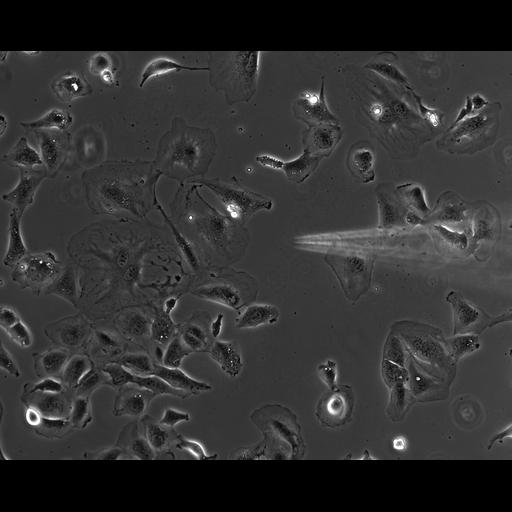
Time lapse movie of mouse embryonic fibroblasts in culture imaged at 30 second intervals by phase contrast microscopy. A micropipette is positioned near the cells to deliver either a growth factor or control buffer. In this sequence PBS buffer is added and the cells move independently of the addition. Also of note, a cell can be seen dividing in the lower left.
These data were collected as research which led to the published article Cell Physiol Biochem 2010;25:279-292. Microscopy was performed with a 10X planapo phase contrast objective on an Olympus IX 70 microscope with a SensicamHQ cooled CCD camera and a heated stage insert set to 37 degrees C. Cells were grown and serum starved for 48 h in a coverslip bottom MatTek dish. Small amounts of PDGF-AA were continuously ejected from the micropipette to create a gradient in the vicinity of growth-arrested wt MEFs. Cell movement was monitored with time lapse video microscopy, taking images at 30 second intervals. A Femtojet Micromanipulator 5171 (Eppendorf-Brinkman Instruments) and a pump (model Femtojet; Eppendorf) were used to control the position of the micropipette and the pressure required for the chemoattractant flow. Femptotip II micropipettes were positioned within 1 μm of the cover slip and pressure set at 30 to 45 hPa. Spatial scale is approximate. Imaging performed at the Image Analysis Facility of the Albert Einstein College of Medicine.
| Spatial Axis | Image Size | Pixel Size |
|---|---|---|
| X | 1280px | 0.33µm |
| Y | 1024px | 0.33µm |
| Time | 30 seconds | 306 |
|---|