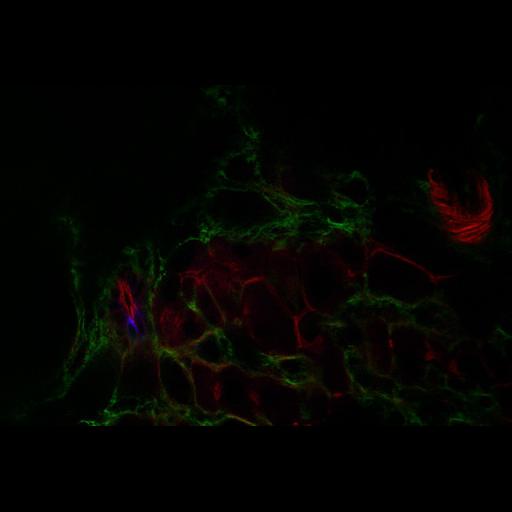
Liver of a rat exposed to chemical carcinogens. Serial sections of the liver imaged by laser scanning confocal microscopy. F-actin (red) is in bile canaliculi at junctions of the hepatocytes, which are the large round cells arranged in a circular mass in the bottom center, in ductule cells, which are above and to the left of the hepatocytes, and in blood vessel endothelial cells, which are in the upper right and far left. Fibronectin (green) is in the tissue outside the hepatocytes. Bile ductule and/or oval cells are marked blue.
The liver of a rat fed a carcinogenic chemicals diet was cryo- and paraformaldehyde-fixed and frozen sections were prepared. The section was labeled with Cy 3 for OV-6, a marker for oval cells/bile ductile cells, Cy5 for fibronectin, and FITC for f-actin. Colors in this image were assigned to each probe by the Cell Imaging Library software. The confocal microscope was a BioRad MRC 600 with a Kr/Ar laser with lines at 488, 568, and 647 nm mounted on a Nikon Diaphot with 60X N.A. 1.4 phase 3 optics. Because the scan head only had two photomultiplier tubes, the filters had to be swapped out manually to collect a series of in register images of more than two fluorescent probes. The microscope was in the Analytical Imaging Facility of the Albert Einstein College of Medicine.
| Spatial Axis | Image Size | Pixel Size |
|---|---|---|
| X | 768px | 0.275µm |
| Y | 512px | 0.275µm |
| Z | 13px | 1µm |