Alternate header for print version


Licensing
Public Domain: This image is in the public domain and thus free of any copyright restrictions. However, as is the norm in scientific publishing and as a matter of courtesy, any user should credit the content provider for any public or private use of this image whenever possible. Learn more
tweet  
*CIL – Cell Image Library accession number. Please use this to reference an image.

CIL:35619*  Cite 
Description

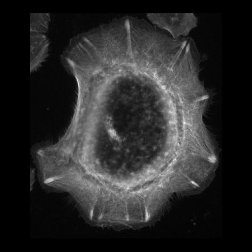
Actin fiber organization in primary keratinocyte. There are numerous actin arcs around the periphery of the cell and actin bundles that terminate in focal adhesions (bright structures perpendicular to edge).

Technical Details

Image collected on a Zeiss Axiovert 200M microscope using a 63X 1.4 NA objective. Image was contrast stretched using Image J.

Biological Sources
NCBI Organism Classification
Homo sapiens
Cell Type
keratinocyte
Cellular Component
actin cytoskeleton
focal adhesion
Biological Context
Biological Process
cytoskeleton organization
Attribution
Names
Ann Wheeler
OTHER
University London, Blizard Institute Advanced Microscopy Facility
Citation
Digital Object Identifier (DOI)
doi:10.7295/W9CIL35619
Archival Resource Key (ARK)
ark:/b7295/w9cil35619
Imaging
Image Type
recorded image
Image Mode
fluorescence microscopy
Parameters Imaged
fluorescence emission
Source of Contrast
distribution of epitope
Visualization Methods
Alexa Fluor 488
phalloidin
Processing History
unprocessed raw data
Sample Preparation
Methods
chemically fixed tissue
Relation To Intact Cell
dispersed cells in vitro
Dimensions
Spatial Axis Image Size Pixel Size
X 390px 0.2µm
Y 457px 0.2µm