Alternate header for print version


Licensing
Attribution Non-Commercial; No Derivatives:This image is licensed under a Creative Commons Attribution, Non-Commercial, No Derivatives License. View License Deed | View Legal Code
tweet  
*CIL – Cell Image Library accession number. Please use this to reference an image.

CIL:35979*  Cite 
Description

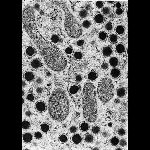
Figure 357 from Chapter 15 (Cytoplasmic Inclusions) of 'The Cell, 2nd Ed.' by Don W. Fawcett M.D. Intestinal epithelium of rat in an advanced stage of fat absorption. In contrast to most cells, lipid droplets in intestinal epithelial cells are bound by membrane. Image from S. Palay and J.P. Revel in Lipid Transport [H.C. Meng, ed.] Charles C. Thomas, Springfield, IL, 1964. A PDF copy of the accompanying chapter is available on the ASCB’s BioEDUCATE website.

Biological Sources
NCBI Organism Classification
Rattus
Cell Type
epithelial cell
Cellular Component
lipid particle
mitochondrion
smooth endoplasmic reticulum
Biological Context
Biological Process
lipid storage
Attribution
Names
Don W. Fawcett
S. Palay; J.P. Revel
Published
ISBN 0721635849
Link
http://www.ascb.org/bioeducate/FawcettTheCell...
Citation
Digital Object Identifier (DOI)
doi:10.7295/W9CIL35979
Archival Resource Key (ARK)
ark:/b7295/w9cil35979
Grouping This image is part of a group.
Imaging
Image Type
transmission electron microscopy (TEM)
Image Mode
illumination by electrons
detection of electrons
Parameters Imaged
electron density
Source of Contrast
stain with broad specificity
Visualization Methods
osmium tetroxide
Data Qualifiers
processed data
Dimensions
Spatial Axis Image Size Pixel Size
X 894px ——
Y 1269px ——