Alternate header for print version


Licensing
Attribution Non-Commercial; No Derivatives:This image is licensed under a Creative Commons Attribution, Non-Commercial, No Derivatives License. View License Deed | View Legal Code
tweet  
*CIL – Cell Image Library accession number. Please use this to reference an image.

CIL:35995*  Cite 
Description

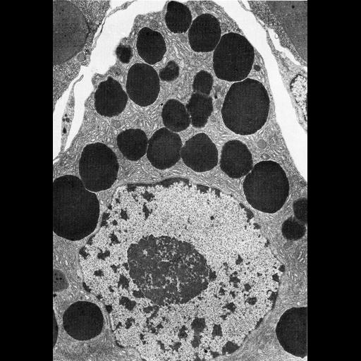
Figure 374 from Chapter 15 (Cytoplasmic Inclusions) of 'The Cell, 2nd Ed.' by Don W. Fawcett M.D. Cell type-f from Acinus III in the salivary gland of the tick Rhipicephalus appendiculatus. These cells are active in synthesis and secretion at the start of a blood meal. A prominent nucleolus, as shown here, is common in cells engaged in synthesis of large amounts of protein for export. A PDF copy of the accompanying chapter is available on the ASCB’s BioEDUCATE website.

Biological Sources
NCBI Organism Classification
Rhipicephalus appendiculatus
Cell Type
glandular epithelial cell
Cellular Component
secretory granule
nucleus
endoplasmic reticulum
nucleolus organizer region
Biological Context
Biological Process
secretory granule organization
Attribution
Names
Don W. Fawcett
Published
ISBN 0721635849
Link
http://www.ascb.org/bioeducate/FawcettTheCell...
Citation
Digital Object Identifier (DOI)
doi:10.7295/W9CIL35995
Archival Resource Key (ARK)
ark:/b7295/w9cil35995
Grouping This image is part of a group.
Imaging
Image Type
transmission electron microscopy (TEM)
Image Mode
illumination by electrons
detection of electrons
Parameters Imaged
electron density
Source of Contrast
stain with broad specificity
Visualization Methods
osmium tetroxide
Data Qualifiers
processed data
Dimensions
Spatial Axis Image Size Pixel Size
X 906px ——
Y 1284px ——