Alternate header for print version


Licensing
Attribution Non-Commercial; No Derivatives:This image is licensed under a Creative Commons Attribution, Non-Commercial, No Derivatives License. View License Deed | View Legal Code
tweet  
*CIL – Cell Image Library accession number. Please use this to reference an image.

CIL:35996*  Cite 
Description

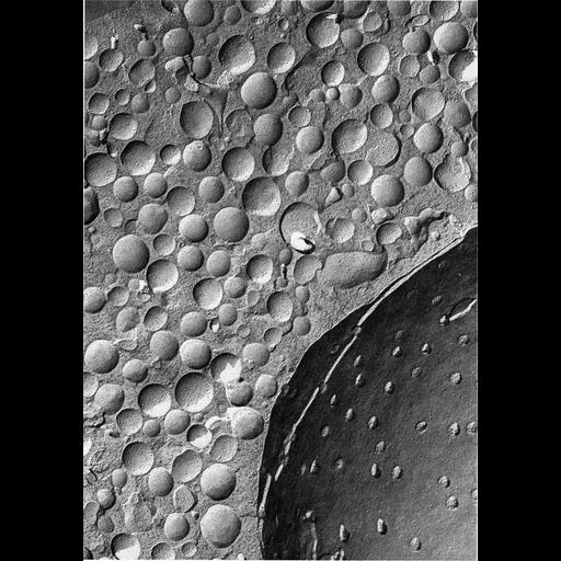
Figure 375 from Chapter 15 (Cytoplasmic Inclusions) of 'The Cell, 2nd Ed.' by Don W. Fawcett M.D. Freeze-fracture replica of a cell from rat adrenal medulla. Using this technique, secretory vesicles appear as smooth-coutoured spheres with intramembraneous particles inserted in the limiting membrane. Nuclear pore complexes are apparent in the fractured inner membrane of the nucleus at the lower right. Image by Daniel Friend. A PDF copy of the accompanying chapter is available on the ASCB’s BioEDUCATE website.

Biological Sources
NCBI Organism Classification
Rattus
Cell Type
adrenal medulla chromaffin cell
Cellular Component
secretory granule
nucleus
nuclear pore
Biological Context
Biological Process
secretory granule organization
Attribution
Names
Don W. Fawcett
Daniel Friend
Published
ISBN 0721635849
Link
http://www.ascb.org/bioeducate/FawcettTheCell...
Citation
Digital Object Identifier (DOI)
doi:10.7295/W9CIL35996
Archival Resource Key (ARK)
ark:/b7295/w9cil35996
Grouping This image is part of a group.
Imaging
Image Type
transmission electron microscopy (TEM)
Image Mode
illumination by electrons
detection of electrons
Parameters Imaged
electron density
Source of Contrast
differences in deposition of metal shadow
Visualization Methods
shadowing and plating
Data Qualifiers
processed data
Sample Preparation
Methods
freeze_fracture/freeze_etch
Relation To Intact Cell
microtome-sectioned tissue
Dimensions
Spatial Axis Image Size Pixel Size
X 897px ——
Y 1275px ——