Alternate header for print version


Licensing
Attribution Non-Commercial; No Derivatives:This image is licensed under a Creative Commons Attribution, Non-Commercial, No Derivatives License. View License Deed | View Legal Code
tweet  
*CIL – Cell Image Library accession number. Please use this to reference an image.

CIL:35998*  Cite 
Description

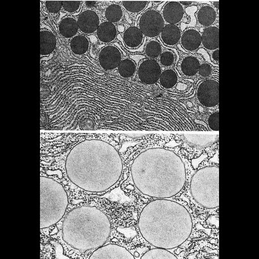
Figures 377 (upper) and 378 (lower) from Chapter 15 (Cytoplasmic Inclusions) of 'The Cell, 2nd Ed.' by Don W. Fawcett M.D. Comparison between the human pancreatic acinar cell (upper) and guinea pig pancreatic cell (lower) reveals species differences in size and density of secretory vesicles in the same cell type. The zymogen granules in the human acinar cell are more dense, or osmiophilic than those of the guinea pig. Upper image by Susumo Ito and Arthur Like; lower from R. Bolender, J. Cell Biol. 61:269-87, 1974. A PDF copy of the accompanying chapter is available on the ASCB’s BioEDUCATE website.

Biological Sources
NCBI Organism Classification
Homo sapiens
Cavia porcellus
Cell Type
acinar cell
Cellular Component
secretory granule
zymogen granule
endoplasmic reticulum
Biological Context
Biological Process
secretory granule organization
Attribution
Names
Don W. Fawcett
Susumo Ito; Arthur Like
Robert Bolender
Published
ISBN 0721635849
Link
http://www.ascb.org/bioeducate/FawcettTheCell...
Citation
Digital Object Identifier (DOI)
doi:10.7295/W9CIL35998
Archival Resource Key (ARK)
ark:/b7295/w9cil35998
Grouping This image is part of a group.
Imaging
Image Type
transmission electron microscopy (TEM)
Image Mode
illumination by electrons
detection of electrons
Parameters Imaged
electron density
Source of Contrast
stain with broad specificity
Visualization Methods
osmium tetroxide
Data Qualifiers
processed data
Dimensions
Spatial Axis Image Size Pixel Size
X 879px ——
Y 1269px ——